
Rough Endoplasmic Reticulum Collection
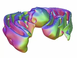
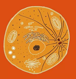
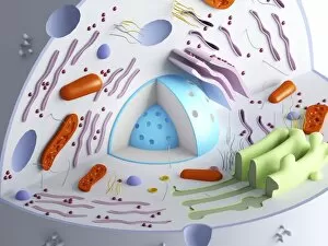
The rough endoplasmic reticulum (RER) is a vital component of the interior of eukaryotic cells

For sale as Licensed Images
Choose your image, Select your licence and Download the media
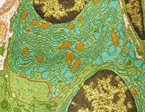
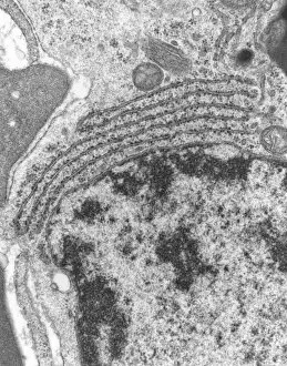
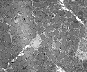
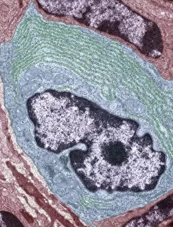
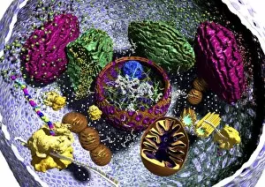
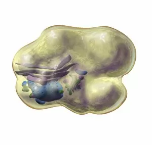
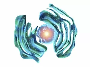
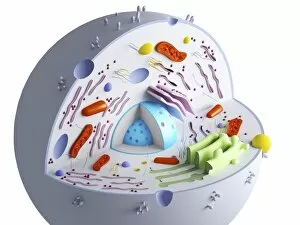
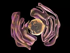
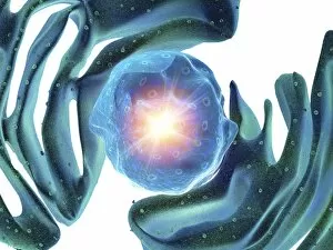
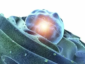
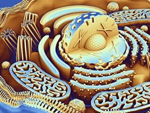
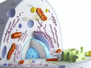
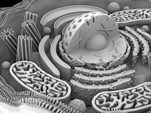
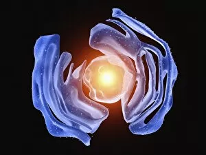
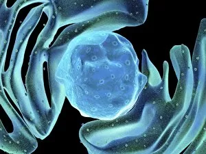
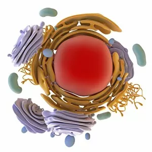
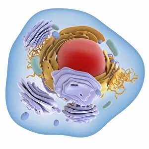
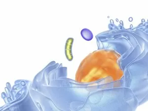
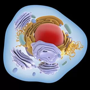
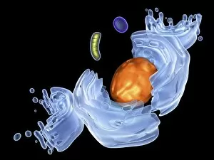
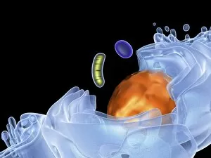
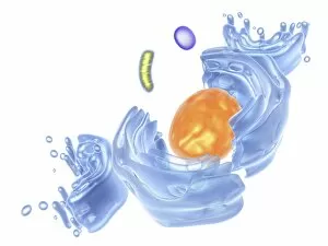
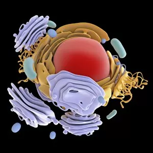
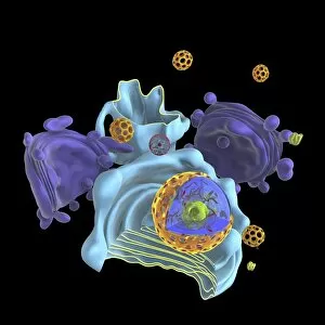
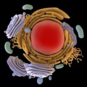
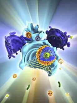
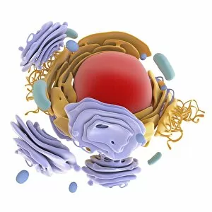
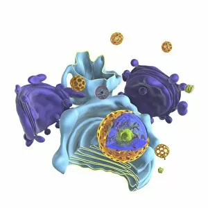
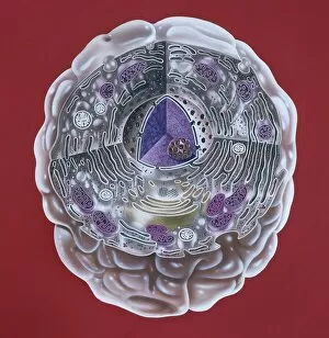
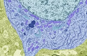
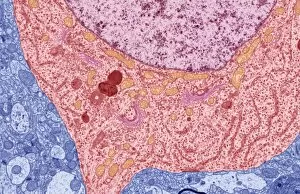
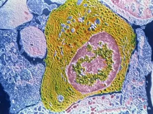
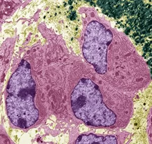
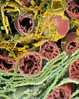
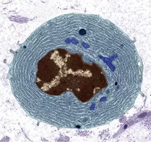
The rough endoplasmic reticulum (RER) is a vital component of the interior of eukaryotic cells. This intricate network of membranes, studded with ribosomes, plays a crucial role in protein synthesis and transport within the cell. Under the powerful lens of a transmission electron microscope (TEM), we can observe the RER's distinctive appearance. The images reveal its close association with the nucleus, forming an interconnected system that ensures efficient communication and coordination between these two essential organelles. In one captivating TEM image, plasma cells are showcased, highlighting their abundant rough endoplasmic reticulum. These specialized immune cells rely on extensive protein production to carry out their critical functions in defending our bodies against pathogens. Further exploration into pancreatic exocrine cells through TEM unravels another fascinating aspect of RER's significance. In pancreatic acinar cells, which secrete digestive enzymes into the small intestine, this organelle dominates their cytoplasm. Its presence emphasizes its pivotal role in synthesizing and packaging proteins destined for secretion. As we delve deeper into these microscopic marvels captured by TEM, it becomes evident that rough endoplasmic reticulum is not just an ordinary cellular structure but rather an intricate web facilitating complex processes within living organisms. From aiding in antibody production to enabling proper digestion through enzyme secretion – all depend on this remarkable organelle's functionality. These mesmerizing glimpses under the microscope offer us a profound appreciation for how intricately designed our cellular machinery truly is. The rough endoplasmic reticulum stands as a testament to nature's brilliance and reminds us that even at such minuscule scales, wonders await those who dare to explore them further.