
Scanning Electron Micrograph Collection (#4)

"Exploring the Microscopic World

Background imageScanning Electron Micrograph Collection: Surface of a rusty screw

Surface of a rusty screw

Background imageScanning Electron Micrograph Collection: Matchstick

Matchstick
Scanning electron microscope (SEM) image showing the fractured surface of a matchstick (x 400 on a standard 9 cm wide print)

Background imageScanning Electron Micrograph Collection: Selaginella kraussiana, spikemoss

Selaginella kraussiana, spikemoss
Scanning electron microscope image of the female spore of Krauss spikemoss (x 150 on a standard 9 cm wide print)

Background imageScanning Electron Micrograph Collection: Hydra sp

Hydra sp
Scanning electron microscope (SEM) image showing the stinging tentacles and mouth of the coelenterate Hydra (x 36 on a standard 9cm wide print)

Background imageScanning Electron Micrograph Collection: Atta cethalotes, leaf-cutter ant

Atta cethalotes, leaf-cutter ant
Scanning electron microscope image of a leaf-cutter ant displayed in the Darwin Centre, at the Natural History Museum, London

Background imageScanning Electron Micrograph Collection: Conodont fossils

Conodont fossils
Scanning electron microscope image of fossils from the Devonian period of northern Estonia, about 465 Ma old ( x 4.2). These creatures are still a mystery to paleontologists

Background imageScanning Electron Micrograph Collection: Small fly, species unknown

Small fly, species unknown
Scanning electron microscope (SEM) of a fly head. Image displayed on the glass screens in the Darwin Centre, at the Natural History Museum, London

Background imageScanning Electron Micrograph Collection: Pelargonium sp. geranium

Pelargonium sp. geranium
Scanning Electron Microscope image of a pelaronium leaf

Background imageScanning Electron Micrograph Collection: Woodlouse antenna

Woodlouse antenna
Scanning Electron Microscope (SEM) image of woodlouse antenna

Background imageScanning Electron Micrograph Collection: Oats

Oats
A scanning electron microscope (SEM) image of oats, artificially coloured by computer

Background imageScanning Electron Micrograph Collection: Spinacia oleracea, spinach

Spinacia oleracea, spinach
A scanning electron microscope (SEM) image of spianch (Spinacia oleracea), artificially coloured by computer

Background imageScanning Electron Micrograph Collection: Solanum sp. tomato

Solanum sp. tomato
A scanning electron microscope (SEM) image of a tomato (Solanum sp.), artificially coloured by computer

Background imageScanning Electron Micrograph Collection: Table salt

Table salt
A scanning electron microscope (SEM) image of table salt, artificially coloured by computer

Background imageScanning Electron Micrograph Collection: Table salt

Table salt
A scanning electron microscope (SEM) image of table salt, artificially coloured by computer

Background imageScanning Electron Micrograph Collection: Oats

Oats
A scanning electron microscope (SEM) image of oats, artificially coloured by computer

Background imageScanning Electron Micrograph Collection: Amphitetras, diatom

Amphitetras, diatom
Scanning electron microscope (SEM) image showing the diatom Amphitetras with its ornate silica shell (x5000 on a standard 9 cm wide print). Coloured artificially by computer

Background imageScanning Electron Micrograph Collection: Vitis sp. red grape

Vitis sp. red grape
A scanning electron microscope (SEM) image of a red grape (Vitis sp.), artificially coloured by computer

Background imageScanning Electron Micrograph Collection: Vitis sp. grape seed

Vitis sp. grape seed
Scanning electron microscope (SEM) image of a grape seed (Vitis sp.), artificially coloured by computer

Background imageScanning Electron Micrograph Collection: Solanum sp. tomato seed

Solanum sp. tomato seed
A scanning electron microscope (SEM) image of a tomato seed (Solanum sp.), artificially coloured by computer

Background imageScanning Electron Micrograph Collection: Leptoglossis ferreyraei

Leptoglossis ferreyraei
A pollen grain of Leptoglossis ferreyraei (polar view) from the family Solanaceae, the tomato family

Background imageScanning Electron Micrograph Collection: Leptoglossis lomana

Leptoglossis lomana
A pollen grain of the Leptoglossis lomana (polar view) from the family Solanacea, the tomato family

Background imageScanning Electron Micrograph Collection: Ciliate plankton

Ciliate plankton
Scanning electron microscope image of a ciliate showing clearly the microscopic hairs or cilia that they use for movement and feeding (x 700)

Background imageScanning Electron Micrograph Collection: Gold in unspecified mineral

Gold in unspecified mineral
Scanning electron microscope image of an elemental map showing the distribution of gold (Au) in mineral samples

Background imageScanning Electron Micrograph Collection: Pyrochlore

Pyrochlore
Scanning electron microscope images of elemental maps showing thorium, uranium, tantalum and silicon in the mineral pyrochlore from Sokli, Finland

Background imageScanning Electron Micrograph Collection: Goyazite

Goyazite
Scanning electron microscope image of the energy-dispersive X-ray spectrum of the mineral goyazite, obtained using Link AN10000 analysis system

Background imageScanning Electron Micrograph Collection: Collembola, springtail

Collembola, springtail
Scanning electron microscope image of a springtail head (x 300)

Background imageScanning Electron Micrograph Collection: Velcro

Velcro
A trademarked name for a fastening tape made up of a strip of nylon with a surface of minute hooks, that fasten to another strip with a surface of uncut pile. A SEM image

Background imageScanning Electron Micrograph Collection: Cells on glass

Cells on glass
Scanning electron microscope (SEM) image of cells on glass (x 2K)

Background imageScanning Electron Micrograph Collection: Volcanic glass, Peles hair

Volcanic glass, Peles hair
Scanning electron microscope image of a sample of volcanic glass from Mt. Pele, produced to evaluate different types of laser in Laser Ablation Inductively Coupled Plasma Mass Spectrometry

Background imageScanning Electron Micrograph Collection: Variable pressure scanning electron microscope

Variable pressure scanning electron microscope
This electron microscope allows the imaging of samples without any preparation

Background imageScanning Electron Micrograph Collection: Myxomycetes, plasmodial slime mould

Myxomycetes, plasmodial slime mould
Scanning electron microscope image of a plasmodial slime mould spore (x12000). This mould spends most of its life as a single cell; when they reproduce they form a slug-like blob that can travel

Background imageScanning Electron Micrograph Collection: Collembola ocelli, springtail

Collembola ocelli, springtail
Scanning electron microscope image of the springtail with simple eyes (x 1.2K)

Background imageScanning Electron Micrograph Collection: Copper in unspecified mineral

Copper in unspecified mineral
Scanning electron microscope image of an elemental map showing the distribution of copper (Cu) in mineral samples

Background imageScanning Electron Micrograph Collection: Ostracod

Ostracod
Scanning electron microscope image of an ostracod, an arthropod where the body is enclosed in a carapace (x 220)

Background imageScanning Electron Micrograph Collection: Collembola sp. springtail

Collembola sp. springtail
Scanning electron microscope image of a springtail showing the characteristic pattern on the cuticle surface (x 3.5K)

Background imageScanning Electron Micrograph Collection: Fractured pollen grain

Fractured pollen grain
Scanning electron microscope (SEM) image showing a fractured pollen grain

Background imageScanning Electron Micrograph Collection: Populus nigra, lombardy or black poplar pollen

Populus nigra, lombardy or black poplar pollen
Scanning electron microscope image (x 1500) of black poplar pollen grains showing a characteristic granular surface ornamentation and no apertures (inaperturate)

Background imageScanning Electron Micrograph Collection: Butterfly wing scale (part)

Butterfly wing scale (part)

Background imageScanning Electron Micrograph Collection: Aphis fabae, black bean aphid

Aphis fabae, black bean aphid
Scanning electron microscope image showing a frontal view of a black bean aphid on leaf (x100). Aphids or plant lice are small, plant-sucking insects

Background imageScanning Electron Micrograph Collection: Human hair

Human hair
Scanning electron microscope (SEM) image showing a human hair with the cuticle reflexed

Background imageScanning Electron Micrograph Collection: Coccolithus pelagicus

Coccolithus pelagicus
Coccosphere of Coccolithus pelagicus, a common cold water coccolithophore. Collected from the British Continental shelf, North West of Scotland. Specimen diameter 15m. False-coloured SEM image

Background imageScanning Electron Micrograph Collection: Ophiaster formosus

Ophiaster formosus
A coccolithophore with long appendages formed of strings of highly modified coccoliths. Collected from the West Pacific. Specimen diameter 50m. False-coloured SEM image

Background imageScanning Electron Micrograph Collection: Lasius niger, black garden ant

Lasius niger, black garden ant

Background imageScanning Electron Micrograph Collection: Calliphora vicina, blowfly or blue bottle

Calliphora vicina, blowfly or blue bottle
Scanning electron microscope (SEM) image of a blowflys wing

Background imageScanning Electron Micrograph Collection: Fractured anther

Fractured anther
Scanning electron microscope (SEM) image showing a fractured anther, otherwise known as the sac, which contains the pollen in the male sex organs (stamens)


For sale as Licensed Images

Choose your image, Select your licence and Download the media

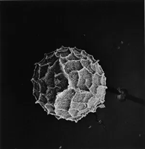
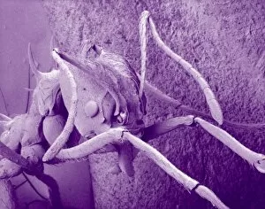
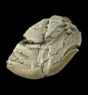
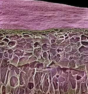
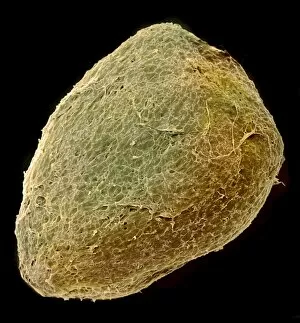
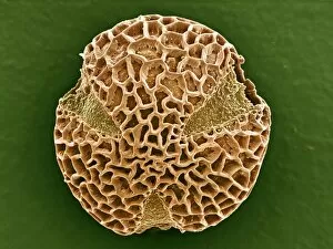
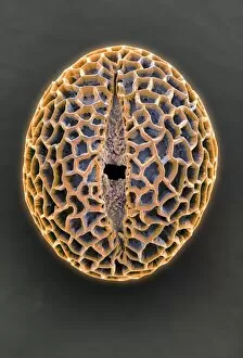
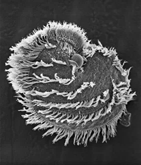
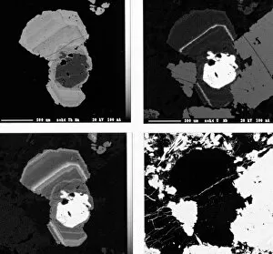
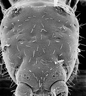
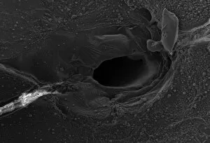
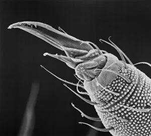
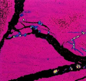
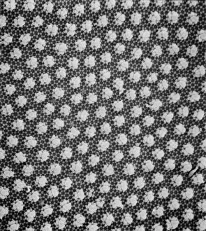
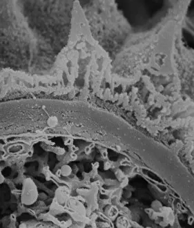
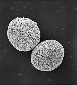
"Exploring the Microscopic World: Captivating Scanning Electron Micrographs" Witness the intricate beauty of nature with this Scanning Electron Micrograph (SEM) capturing a Praying Mantis in stunning detail, magnified 30 times its size. Delve into the microscopic realm and discover the resilience of Tardigrades, also known as 'Water Bears, ' through this SEM image magnified at an astounding 1250 times. Uncover the hidden dangers lurking within Crysotile asbestos as revealed by this powerful SEM, shedding light on its harmful effects on human health. Journey inside the liver through a fascinating SEM, offering a glimpse into its complex structure and vital role in our body's functions. Get up close and personal with Cimex lectularius, commonly known as bed bugs, thanks to this detailed SEM that exposes their tiny yet formidable presence. Marvel at the intricate features of a Fruit Fly captured under an SEM lens at 300 times magnification – revealing astonishing details invisible to the naked eye. Explore Taraxacum officinale or dandelion's fruiting head like never before with this captivating SEM image showcasing its delicate structures and unique design. Discover nature's engineering marvels by examining Snail teeth under high-powered microscopy; witness how these minuscule structures aid them in feeding and survival. Peer into another world within Plasmodium sp. , a malarial parasite responsible for widespread disease transmission worldwide – all unveiled through this striking SEM image. Dive deep into geology with Kaolinite - explore its fine crystalline structure using an advanced scanning electron microscope to unravel its mineralogical secrets. Encounter Sarcoptes scabiei or scabies mite up close via an extraordinary SEM capture; gain insight into their morphology while appreciating their impact on human health.