Scanning Electron Micrograph Collection (#6)

"Exploring the Microscopic World

Background imageScanning Electron Micrograph Collection: Asteraceae, daisy

Asteraceae, daisy
Scanning electron microscope image of the fractured surface of an anther showing a developing pollen grain from a member of the daisy or Asteraceae family ( X 3000)

Background imageScanning Electron Micrograph Collection: Lumbricus terrestris, earthworm

Lumbricus terrestris, earthworm
Scanning electron microscope (SEM) image showing the chaeta/setae - involved in the locomotion on an earthworm

Background imageScanning Electron Micrograph Collection: Taraxacum officinale, dandelion

Taraxacum officinale, dandelion
Scanning electron microscope (SEM) image of a dandelion (x 80)

Background imageScanning Electron Micrograph Collection: Spider trichobothrium hair

Spider trichobothrium hair
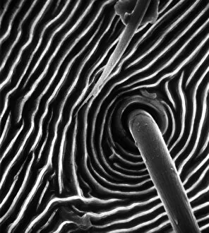
Scanning electron microscope (SEM) image of the base of a trichobothrium hair (x 1, 000). The hair is an air-movement sensor extending from the pit in the cuticle of a spiders leg

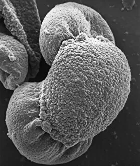
Background imageScanning Electron Micrograph Collection: Fagus sylvatica, European beech pollen

Fagus sylvatica, European beech pollen
Scanning electron microscope picture (X1500) showing a pollen grain as seen from the side. The image shows one of the three laterally-placed aperture furrows with a small pore in the centre

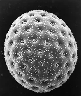
Background imageScanning Electron Micrograph Collection: Fraxinus excelsior, weeping ash pollen

Fraxinus excelsior, weeping ash pollen
Scanning electron microscope picture (x 1500) of ash pollen grains from above, with three furrowed apertures (trizonocolporate)

Background imageScanning Electron Micrograph Collection: Calyptrolithophora papillifera, holococcolith

Calyptrolithophora papillifera, holococcolith
An SEM of a holococcolith, a nano-fossil, with flat top

Background imageScanning Electron Micrograph Collection: A bryozoan colony

A bryozoan colony
Scanning electron microscope image displayed on the glass screens in the Darwin Centre, at the Natural History Museum, London

Background imageScanning Electron Micrograph Collection: Cystopteris diaphana, diaphanous bladder fern

Cystopteris diaphana, diaphanous bladder fern
An SEM showing a close-up of the spiny-lacunar surface of the diaphanous bladder fern (Cystopteris diaphana) spore. Photographed using Philips XL30 SEM

Background imageScanning Electron Micrograph Collection: Hair of the Dog

Hair of the Dog
A scanning electron micrograph (SEM) of a dog hair

Background imageScanning Electron Micrograph Collection: Mustelus mustelus, smoothhound shark

Mustelus mustelus, smoothhound shark
A Scanning Electron Microscope image of smoothhound shark skin. The skin is covered with tiny teeth called dermal denticles

Background imageScanning Electron Micrograph Collection: Fragaria sp. strawberry

Fragaria sp. strawberry
A scanning electron microscope (SEM) image of a strawberry (Fragaria sp.), artificially coloured by computer

Background imageScanning Electron Micrograph Collection: Sugar grains

Sugar grains
A scanning electron microscope (SEM) image of sugar grains, artificially coloured by computer

Background imageScanning Electron Micrograph Collection: Vitis sp. white grape

Vitis sp. white grape
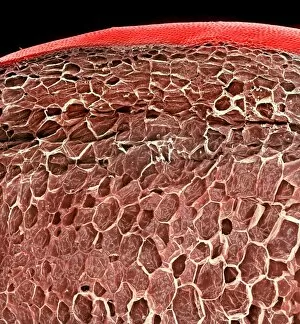
A scanning electron microscope (SEM) image of a white grape (Vitis sp.), artificially coloured by computer

Background imageScanning Electron Micrograph Collection: Browallia speciosa, amethyst

Browallia speciosa, amethyst
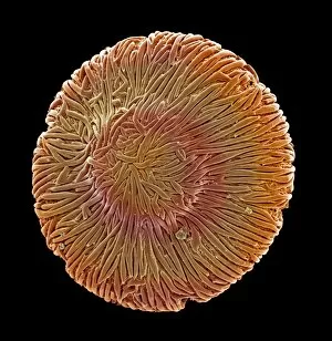
A pollen grain of the Browallia speciosa (polar view) from the family Solanaceae, the tomato family

Background imageScanning Electron Micrograph Collection: Vaccinium sp. blueberry

Vaccinium sp. blueberry
A scanning electron microscope (SEM) image of a blueberry (Vaccinium sp.), artificially coloured by computer

Background imageScanning Electron Micrograph Collection: Solanum sp. tomato

Solanum sp. tomato
A scanning electron microscope (SEM) image of a tomato (Solanum sp.), artificially coloured by computer

Background imageScanning Electron Micrograph Collection: Isurus oxyrinchus, mako shark

Isurus oxyrinchus, mako shark
Scanning Electron Microscope image of mako shark skin

Background imageScanning Electron Micrograph Collection: Lasius niger, black garden ant

Lasius niger, black garden ant
Scanning electron microscope (SEM) of a black ant leg. Widespread and common in a range of habitats but perhaps most familiar in gardens where nests are formed under paving stones and brickwork

Background imageScanning Electron Micrograph Collection: Porcellio sccaber, woodlouse

Porcellio sccaber, woodlouse
Scanning electron microscope (SEM) image showing all the units that make up the compound eye of a woodlouse

Background imageScanning Electron Micrograph Collection: Calliphora vicina, blowfly or bluebottle

Calliphora vicina, blowfly or bluebottle
Scanning electron microscope (SEM) image of a blowfly proboscis (x 85). This specialised mouth-part is used to squirt digestive enzymes onto the food

Background imageScanning Electron Micrograph Collection: Papilio machaon, old world swallowtail

Papilio machaon, old world swallowtail
SEM image of a Papilio machaon wing

Background imageScanning Electron Micrograph Collection: Danionella dracula, minnow

Danionella dracula, minnow
SEM image of the Danionella dracula. This tiny 17mm fish has evolved many unique and unusual characteristics, the most spectacular of which are jaw modifications that resemble true teeth

Background imageScanning Electron Micrograph Collection: Feather detail

Feather detail

Background imageScanning Electron Micrograph Collection: Pollen on bee

Pollen on bee
Scanning electron microscope (SEM) image of pollen on a bee. If the plant depends on animals for pollination, the pollen will be relatively large and sticky

Background imageScanning Electron Micrograph Collection: Dermestes lardarius, larder beetle

Dermestes lardarius, larder beetle
Scanning electron microscope image of a larder beetle (x22). These beetles are important for the damage they do, mainly through feeding on animal matter. Coloured artificially by computer

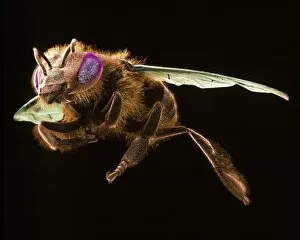
Background imageScanning Electron Micrograph Collection: Apis mellifera, honey bee

Apis mellifera, honey bee
Scanning electron microscope image of a honey bee coloured artificially by computer. The female worker caste of this species have special baskets on their legs to to take pollen back to the nest

Background imageScanning Electron Micrograph Collection: Actinopora disticha, bryozoan

Actinopora disticha, bryozoan
Scanning electron micrograph of a fossil cyclostome bryozoan from the Cretaceous Chalk, Santonian, Kent

Background imageScanning Electron Micrograph Collection: Wilbertopora woodwardi (Brydone), bryozoan

Wilbertopora woodwardi (Brydone), bryozoan
Scanning electron micrograph of a fossil cheilostome bryozoan. Specimen originates from the Upper Cretaceous Chalk, West Mean Station, Hampshire, U.K

Background imageScanning Electron Micrograph Collection: Ptinus tectus, spider beetle

Ptinus tectus, spider beetle
Scanning electron microscope image of a spider beetle (x 9). The long antennae, hairy body and waist-like constriction give this beetle the appearance of a spider

Background imageScanning Electron Micrograph Collection: Aspidelectra melolontha, bryozoan

Aspidelectra melolontha, bryozoan
Scanning electron micrograph. Zooids of a bleached colony of a modern cheilostome bryozoan. A recent specimen from Sheppey, Kent

Background imageScanning Electron Micrograph Collection: Pinus sylvestris, scots pine

Pinus sylvestris, scots pine
Scanning electron microscope (SEM) image showing a pollen grain from a scots pine. Note the air bladders that help it to float through the air (x 1500 on a standard 9 cm wide print)

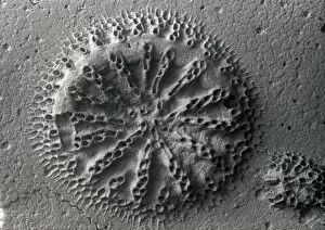
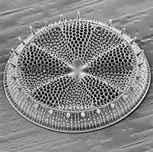
Background imageScanning Electron Micrograph Collection: Actinoptychus, diatom

Actinoptychus, diatom
Scanning electron microscope image of the exterior valve of the diatom Actinoptychus (x 500 on a standard 9 cm wide print)

Background imageScanning Electron Micrograph Collection: Chenopodium album, goosefoot

Chenopodium album, goosefoot
Scanning electron microscope image of a pollen grain from a member of the goosefoot family (x 3000 on a standard 9 cm wide print)

Background imageScanning Electron Micrograph Collection: Marine Diatoms and Radiolarians

Marine Diatoms and Radiolarians
LRMC-17 Scanning Electron Micrograph (SEM): Marine Diatoms and Radiolaria skeletons Magnification x435 (when printed A4, 29.7 cm cm wide)

Background imageScanning Electron Micrograph Collection: Scanning Electron Micrograph (SEM): Fusarium, Magnification x 1, 600 (A4 size: 29. 7 cm width)

Scanning Electron Micrograph (SEM): Fusarium, Magnification x 1, 600 (A4 size: 29. 7 cm width)
LRDS-60 Fusarium Scanning Electron Micrograph (SEM) Fusarium graminearum Magnification x 1, 600 (A4 size: 29.7 cm width) Coloured by hand to enhance natural features

Background imageScanning Electron Micrograph Collection: Scanning Electron Micrograph (SEM): E. coli; Magnification x 15, 000 (A4 size: 29. 7 cm width)

Scanning Electron Micrograph (SEM): E. coli; Magnification x 15, 000 (A4 size: 29. 7 cm width)
LRDS-40 E. coli Scanning Electron Micrograph (SEM) Escherichia coli Magnification x 15, 000 (A4 size: 29.7 cm width) Coloured by hand to enhance natural features

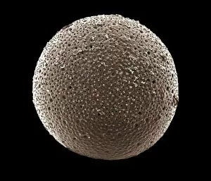
Background imageScanning Electron Micrograph Collection: Foraminifera

Foraminifera
LRDS-306 Scanning Electron Micrograph (SEM): Foraminifera Magnification x400 (when printed A4 size, 29.7 cm wide) Orbulina universa David Spears (Last Refuge) / ardea.com Last Refuge contact details

Background imageScanning Electron Micrograph Collection: Marine Diatom

Marine Diatom
LRDS-293 Scanning Electron Micrograph (SEM): Marine Diatom Magnification x2, 570 (when printed 10.5 cm wide) Bacteriastrum sp David Spears (Last Refuge) / ardea.com Last Refuge contact details


For sale as Licensed Images

Choose your image, Select your licence and Download the media

"Exploring the Microscopic World: Captivating Scanning Electron Micrographs" Witness the intricate beauty of nature with this Scanning Electron Micrograph (SEM) capturing a Praying Mantis in stunning detail, magnified 30 times its size. Delve into the microscopic realm and discover the resilience of Tardigrades, also known as 'Water Bears, ' through this SEM image magnified at an astounding 1250 times. Uncover the hidden dangers lurking within Crysotile asbestos as revealed by this powerful SEM, shedding light on its harmful effects on human health. Journey inside the liver through a fascinating SEM, offering a glimpse into its complex structure and vital role in our body's functions. Get up close and personal with Cimex lectularius, commonly known as bed bugs, thanks to this detailed SEM that exposes their tiny yet formidable presence. Marvel at the intricate features of a Fruit Fly captured under an SEM lens at 300 times magnification – revealing astonishing details invisible to the naked eye. Explore Taraxacum officinale or dandelion's fruiting head like never before with this captivating SEM image showcasing its delicate structures and unique design. Discover nature's engineering marvels by examining Snail teeth under high-powered microscopy; witness how these minuscule structures aid them in feeding and survival. Peer into another world within Plasmodium sp. , a malarial parasite responsible for widespread disease transmission worldwide – all unveiled through this striking SEM image. Dive deep into geology with Kaolinite - explore its fine crystalline structure using an advanced scanning electron microscope to unravel its mineralogical secrets. Encounter Sarcoptes scabiei or scabies mite up close via an extraordinary SEM capture; gain insight into their morphology while appreciating their impact on human health.