Scanning Electron Microscope Collection (#100)

"Unveiling the Hidden World

Background imageScanning Electron Microscope Collection: Planktonic diatom alga, SEM

Planktonic diatom alga, SEM
Planktonic diatom alga. Coloured scanning electron micrograph (SEM) of a diatom alga. A diatom is a planktonic unicellular alga that has silica in its cell wall (frustule)

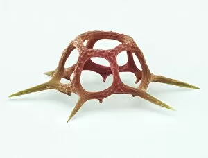
Background imageScanning Electron Microscope Collection: Silicoflagellate, SEM

Silicoflagellate, SEM
Silicoflagellate. Coloured scanning electron micrograph (SEM) of the skeleton of a Dictyocha speculum silicoflagellate. Silicoflagellates are single-celled marine algae

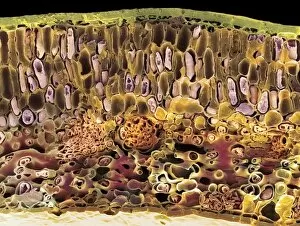
Background imageScanning Electron Microscope Collection: Sago cycad leaf, SEM

Sago cycad leaf, SEM
Sago cycad leaf. Coloured scanning electron micrograph (SEM) of a section through a sago cycad (Cycas revoluta) leaf. At lower centre and left are vascular bundles (orange clusters)

Background imageScanning Electron Microscope Collection: Flowering fern spores, SEM

Flowering fern spores, SEM
Flowering fern spores. Coloured scanning electron micrograph (SEM) of three spores of the flowering fern (Osmunda regalis). The flowering, or royal, fern, is found across much of the world

Background imageScanning Electron Microscope Collection: Escherichia coli bacteria, SEM

Escherichia coli bacteria, SEM
Escherichia coli bacteria, coloured scanning electron micrograph (SEM). E. coli bacteria are a normal part of the intestinal flora in humans and other animals, where they aid digestion

Background imageScanning Electron Microscope Collection: Salmonella typhimurium bacteria, SEM

Salmonella typhimurium bacteria, SEM
Salmonella typhimurium bacteria, coloured scanning electron micrograph (SEM). These bacteria, also known as Salmonella enterica serovar typhimurium, are Gram-negative

Background imageScanning Electron Microscope Collection: Marine diatom alga, SEM

Marine diatom alga, SEM
Diatom alga. Coloured scanning electron micrograph (SEM) of part of a marine diatom alga. Diatoms form a large part of the marine phytoplankton

Background imageScanning Electron Microscope Collection: Freshwater alga, SEM

Freshwater alga, SEM
Freshwater alga. Coloured scanning electron micrograph (SEM) of a single-celled freshwater alga

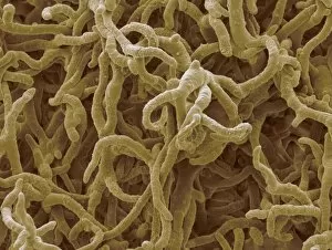
Background imageScanning Electron Microscope Collection: Fungal hyphae, SEM

Fungal hyphae, SEM
Fungal hyphae. Coloured scanning electron micrograph (SEM) of the hyphae of a fungus. Hyphae are thread-like structures that form the mycelium

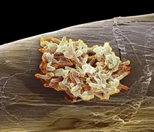
Background imageScanning Electron Microscope Collection: Trichophyton fungus, SEM

Trichophyton fungus, SEM
Trichophyton fungus. Coloured scanning electron micrograph (SEM) of a dermatophyte fungus belonging to the genus Trichophyton

Background imageScanning Electron Microscope Collection: MRSA bacteria, SEM

MRSA bacteria, SEM
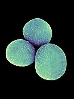
MRSA bacteria, coloured scanning electron micrograph (SEM). Methicillin-resistant Staphylococcus aureus (MRSA) is a Gram-positive, round (coccus) bacterium

Background imageScanning Electron Microscope Collection: Staphylococcus aureus bacteria, SEM

Staphylococcus aureus bacteria, SEM
Staphylococcus aureus bacteria, coloured scanning electron micrograph (SEM). These Gram-positive cocci (round) are found on the skin and mucous membranes of humans and many animals

Background imageScanning Electron Microscope Collection: Bread mould

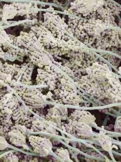
Bread mould (Penicillium sp.). Coloured scanning electron micrograph (SEM) of mouldy bread. The fungus penetrates the bread, digesting and absorbing its nutrients


For sale as Licensed Images

Choose your image, Select your licence and Download the media

"Unveiling the Hidden World: Exploring with a Scanning Electron Microscope" The scanning electron microscope (SEM) is an incredible tool that allows us to delve into the microscopic realm, revealing intricate details and unlocking secrets of the unseen. With its high magnification capabilities, it offers a glimpse into fascinating aspects of life and nature. In one captivating image, we witness the beauty of gender determination as X and Y chromosomes are captured under the SEM's lens. The intricate patterns and structures within these chromosomes showcase the complexity of our genetic makeup. Moving on to another astonishing discovery, a Scanning Electron Micrograph presents us with an up-close view of a Praying Mantis at 30 times magnification. Every tiny detail becomes visible, from its delicate limbs to its mesmerizing eyes – reminding us of nature's remarkable diversity. Shifting focus to plant life, lavender pollen grains come alive in another SEM image. These minuscule particles take center stage as their unique shapes and textures become apparent under intense scrutiny. Zooming in even further reveals a Tardigrade or 'Water Bear' at an astounding 1250 times magnification. This resilient creature captures our imagination as we explore its alien-like features through this powerful imaging technique. Delving deeper into biological wonders, T lymphocytes battling cancer cells are showcased in yet another breathtaking SEM capture. This visual representation highlights the ongoing fight within our bodies against diseases like cancer – emphasizing both hope and resilience. Calcareous phytoplankton takes center stage next as we uncover their intricate forms through SEM imagery. These microscopic organisms play crucial roles in marine ecosystems while leaving us awe-inspired by their stunningly detailed structures. Transitioning to neural marvels, nerve cells reveal their intricacy under the watchful eye of an SEM. Their branching extensions create complex networks that enable communication throughout our bodies – showcasing nature's ingenuity at work. Venturing into insect realms once again, a fruit fly is immortalized under the SEM's lens.