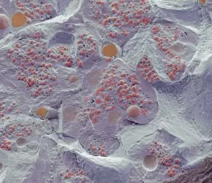
Secretory Collection (#2)

"Unveiling the Hidden Secrets: Exploring the Intricacies Cells and Glands" Delving deep into the intricate workings of our body

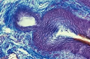
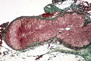
Background imageSecretory Collection: Sebaceous gland, light micrograph C015 / 6911

Sebaceous gland, light micrograph C015 / 6911
Sebaceous gland. Light micrograph of a section through breast tissue showing a sebaceous gland. These glands produce an oily protective substance called sebum

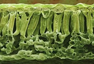
Background imageSecretory Collection: Potato Leaf ts (SEM)

Potato Leaf ts (SEM)
Potato leaf. Coloured scanning electron micrograph (SEM) of a transverse section of a potato leaf (Solanum tuberosum). The upper

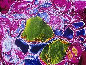
Background imageSecretory Collection: Thyroid gland, SEM C015 / 6419

Thyroid gland, SEM C015 / 6419
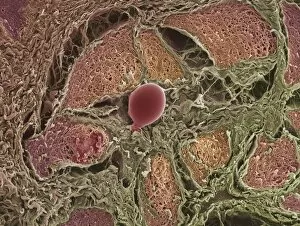
Thyroid gland. Coloured scanning electron micrograph (SEM) of a fracture through the thyroid gland revealing several follicles (green and blue). Between the follicles is connective tissue (pink)

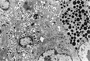
Background imageSecretory Collection: Islet of Langerhans, TEM C015 / 6418

Islet of Langerhans, TEM C015 / 6418
Islet of Langerhans. Transmission electron micrograph (TEM) of a section through cells clustered in an islet of Langerhans in the pancreas

Background imageSecretory Collection: Adrenal cortex, light micrograph C015 / 6417

Adrenal cortex, light micrograph C015 / 6417
Adrenal cortex. Light micrograph of a section through a healthy adrenal cortex (red). The adrenal glands are found on top of the kidneys

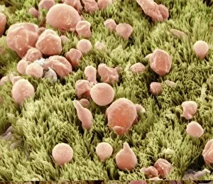
Background imageSecretory Collection: Trachea lining, SEM C013 / 7127

Trachea lining, SEM C013 / 7127
Trachea lining. Coloured scanning electron micrograph (SEM) of the lining of a trachea (wind pipe), showing the cilia (hair-like) and goblet cells (pink). The trachea links the larynx to the lungs

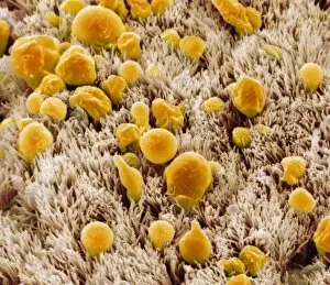
Background imageSecretory Collection: Trachea lining, SEM C013 / 7125

Trachea lining, SEM C013 / 7125
Trachea lining. Coloured scanning electron micrograph (SEM) of the lining of a trachea (wind pipe), showing the cilia (hair-like) and goblet cells (orange). The trachea links the larynx to the lungs

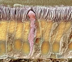
Background imageSecretory Collection: Trachea lining, SEM C013 / 7124

Trachea lining, SEM C013 / 7124
Trachea lining. Coloured scanning electron micrograph (SEM) of a section through the wall of a trachea (wind pipe). The trachea links the larynx to the lungs

Background imageSecretory Collection: Trachea lining, SEM C013 / 7123

Trachea lining, SEM C013 / 7123
Trachea lining. Coloured scanning electron micrograph (SEM) of a section through the wall of a trachea (wind pipe). The trachea links the larynx to the lungs

Background imageSecretory Collection: Lavender leaf oil gland, SEM

Lavender leaf oil gland, SEM
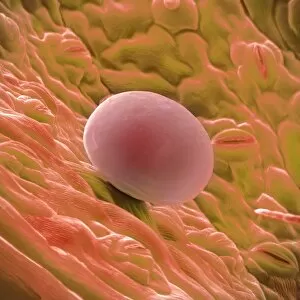
Lavender leaf oil gland. Coloured scanning electron micrograph (SEM) of an oil gland on the surface of a French lavender (Lavandula dentata) leaf

Background imageSecretory Collection: Sage leaf oil gland

Sage leaf oil gland. Coloured scanning electron micrograph (SEM) of a freeze-fracture section through a leaf of sage, Salvia officinalis, showing an oil gland. At lower frame are leaf cells

Background imageSecretory Collection: Sage leaf oil glands

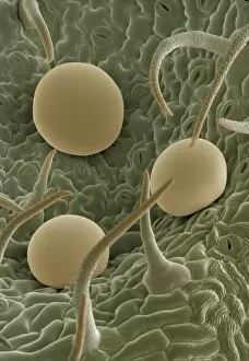
Sage leaf oil glands. Coloured scanning electron micrograph (SEM) of hairs and glands on a leaf of the herb sage, Salvia officinalis. On the leaf surface are two types of hairs, called trichomes

Background imageSecretory Collection: Venus flytrap digestive glands, SEM

Venus flytrap digestive glands, SEM
Venus flytrap digestive glands. Coloured scanning electron micrograph (SEM) of digestive glands from a Venus flytrap plant (Dionaea muscipula)

Background imageSecretory Collection: Sundew carnivorous plant leaf, SEM

Sundew carnivorous plant leaf, SEM
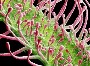
Sundew leaf. Coloured scanning electron micrograph (SEM) of the sticky stalks (red) on the leaf of a cape sundew (Drosera capensis), a carnivorous plant

Background imageSecretory Collection: Potato leaf, SEM

Potato leaf, SEM
Potato leaf. Coloured scanning electron micrograph (SEM) of the surface of a potato leaf (Solanum tuberosum). It is covered in hair-like growths called trichomes

Background imageSecretory Collection: Sweat gland in skin

Sweat gland in skin. Light micrograph of a section through the dermis of the skin showing a sweat gland. The gland is composed of a secretory portion (upper left)

Background imageSecretory Collection: Hair follicles, SEM

Hair follicles, SEM
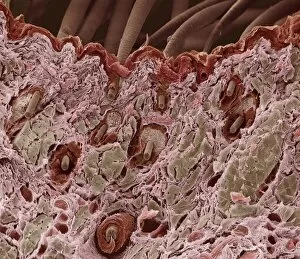
Hair follicles. Coloured scanning electron micrograph (SEM) of a section through freeze- fractured hair follicles in the skin

Background imageSecretory Collection: Thyroid gland follicle, TEM

Thyroid gland follicle, TEM
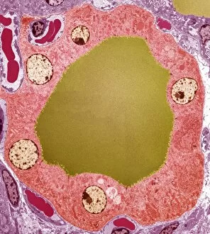
Thyroid gland follicle. Coloured transmission electron micrograph (TEM) of a follicle from the thyroid gland. The follicle consists of a layer of epithelial cells (pink)

Background imageSecretory Collection: Corpus luteum, SEM

Corpus luteum, SEM
Corpus luteum. Coloured scanning electron micrograph (SEM) of a corpus luteum, or yellow body (brown), in an ovary. The corpus luteum is formed after ovulation

Background imageSecretory Collection: Pancreas cell, TEM

Pancreas cell, TEM
Pancreas cell. Coloured transmission electron micrograph (TEM) of an acinar (exocrine) pancreatic cell. Acinar cells secrete the inactive precursors (zymogens)

Background imageSecretory Collection: Small intestine villus, SEM

Small intestine villus, SEM
Small intestine villus. Coloured scanning electron micrograph (SEM) of a freeze fracture section through a villus from the mucosal lining of the small intestine

Background imageSecretory Collection: Trachea lining, TEM

Trachea lining, TEM
Trachea lining. Coloured transmission electron micrograph (TEM) of a transverse section through the lining of the trachea (windpipe). The trachea links the larynx to the lungs

Background imageSecretory Collection: Internal wall of uterus, SEM

Internal wall of uterus, SEM
Internal wall of uterus. Coloured scanning electron micrograph of the mucosa, also known as endometrium, lining the body of the human uterus

Background imageSecretory Collection: Brunners glands in the duodenum

Brunners glands in the duodenum
Brunners glands. Fluorescence confocal light micrograph of a section through a human duodenum (small intestine) showing Brunners glands (blue)

Background imageSecretory Collection: Secretory cells in pancreas, SEM

Secretory cells in pancreas, SEM
Pancreatic secretory cells. Coloured scanning electron micrograph (SEM) of a freeze-fracture through a healthy pancreas, showing the secretory tissue

Background imageSecretory Collection: Small intestine lining, SEM

Small intestine lining, SEM
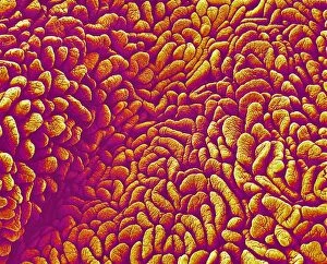
Small intestine villi. Coloured scanning electron micrograph (SEM) of villi (folds) on the lining of the small intestine. Villi greatly increase the intestinal surface area for absorbing nutrients

Background imageSecretory Collection: Small intestine villi, SEM

Small intestine villi, SEM
Small intestine villi. Coloured scanning electron micrograph (SEM) of villi (folds) on the lining of the small intestine. Villi greatly increase the intestinal surface area for absorbing nutrients

Background imageSecretory Collection: Lactating breast tissue, SEM

Lactating breast tissue, SEM

Background imageSecretory Collection: Colon lining

Colon lining. Fluorescence confocal light micrograph of the lining of a mouse colon (large intestine). The colon starts at the small intestine and ends at the rectum

Background imageSecretory Collection: Lacrimal gland, SEM

Lacrimal gland, SEM

Background imageSecretory Collection: Salivary gland, light micrograph

Salivary gland, light micrograph
Salivary gland. Coloured light micrograph of a section through a sublingual salivary gland, which is situated directly under the tongue

Background imageSecretory Collection: Macrophage white blood cell, SEM

Macrophage white blood cell, SEM
Macrophage. Coloured scanning electron micrograph (SEM) of a macrophage white blood cell at the site of a skin wound. Macrophages are cells of the bodys immune system

Background imageSecretory Collection: Fallopian tube, TEM

Fallopian tube, TEM
Fallopian tube, coloured transmission electron micrograph (TEM). Section through non-ciliated columnar epithelium from a fallopian tube. The ciliated cells are not shown here

Background imageSecretory Collection: Trachea mucous membrane, SEM

Trachea mucous membrane, SEM
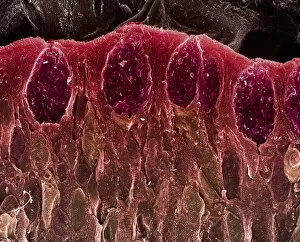
Trachea mucous membrane. Coloured scanning electron micrograph (SEM) of a fractured mucous membrane of the trachea (wind pipe), showing the epithelium and underlying connective tissue

Background imageSecretory Collection: Fallopian tube cells, SEM

Fallopian tube cells, SEM
Fallopian tube cells. Coloured scanning electron micrograph (SEM) of the surface of a fallopian tube, showing non-ciliated (pink) and ciliated (yellow) cells

Background imageSecretory Collection: Marjoram leaf surface, SEM

Marjoram leaf surface, SEM
Marjoram (Origanum vulgare) leaf surface, coloured scanning electron micrograph (SEM). Glandular trichomes are yellow, hair-like trichomes are blue

Background imageSecretory Collection: Pancreas tissue, SEM

Pancreas tissue, SEM
Pancreas tissue. Coloured scanning electron micrograph (SEM) of fractured pancreas tissue, showing numerous acinar cells, containing secretory zymogen granules


For sale as Licensed Images

Choose your image, Select your licence and Download the media

"Unveiling the Hidden Secrets: Exploring the Intricacies Cells and Glands" Delving deep into the intricate workings of our body, we uncover a world hints that hold immense significance. From the medulla oblongata in the brain to osteocyte bone cells, each structure plays a vital role in maintaining our well-being. Intriguing artwork showcases the complexity of these secretory systems. The medulla oblongata, often referred to as the "brain's control center, " orchestrates various bodily functions with its network of neurons. Meanwhile, osteocyte bone cells reveal their delicate intricacy under SEM C016 / 9025, highlighting their crucial role in bone formation and maintenance. Moving on to another remarkable organ, we encounter an artistic representation of pancreas anatomy. This gland houses pancreatic islets of Langerhans - clusters of specialized cells responsible for secreting hormones like insulin and glucagon that regulate blood sugar levels. Venturing further into microscopic realms, TEM unveils plasma cells - guardians against infections by producing antibodies that defend our immune system. Thyme leaf oil glands capture attention with their unique secretion properties while contributing to aromatic experiences. SEM images offer glimpses into hidden worlds within us - brain lining displaying its intricate web-like structure; fallopian tube cells showcasing their elegant arrangement; nasal lining revealing its finely textured surface; cervix exhibiting distinctive features under light micrograph F006 / 9805; trachea lining demonstrating its protective capabilities through SEM C013 / 7126. These captivating visualizations remind us of the awe-inspiring complexity present within our bodies' secretory systems. They serve as a testament to nature's ingenuity and inspire us to delve deeper into understanding these hidden secrets that contribute so profoundly to our overall health and well-being.