Secretory Collection (#4)

"Unveiling the Hidden Secrets: Exploring the Intricacies Cells and Glands" Delving deep into the intricate workings of our body

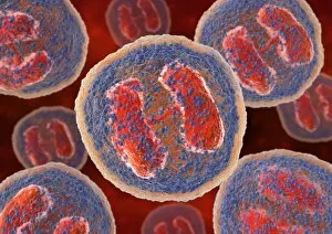
Background imageSecretory Collection: Basophil white blood cells, artwork

Basophil white blood cells, artwork
Basophil white blood cells. Computer artwork of basophil white blood cells, showing their lobed nuclei (red). Basophils are the smallest and least common of the white blood cells

Background imageSecretory Collection: Frog skin glands, light micrograph

Frog skin glands, light micrograph
Frog skin glands. Light micrograph of a section through the skin of a frog, showing secretory glands (round, upper centre) just below the surface (across top)

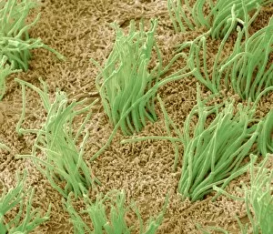
Background imageSecretory Collection: Brain lining, SEM

Brain lining, SEM
Brain lining. Coloured scanning electron micrograph (SEM) of the lining of the brain, showing the ciliary hairs (green) of ependymal cells

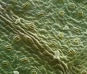
Background imageSecretory Collection: Mint leaf surface, SEM

Mint leaf surface, SEM
Mint leaf surface. Coloured scanning electron micrograph (SEM) of the surface of a mint (Menta sp.) leaf. The white structures are oil glands

Background imageSecretory Collection: Fallopian tube, SEM

Fallopian tube, SEM
Fallopian tube. Coloured scanning electron micrograph (SEM) of the lining of a fallopian tube (oviduct). The fallopian tubes carry the egg from the ovary to the uterus (womb)

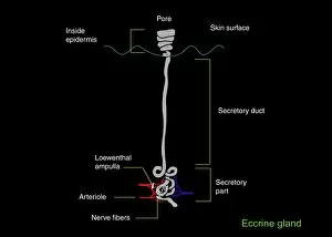
Background imageSecretory Collection: Eccrine sweat gland, artwork

Eccrine sweat gland, artwork
Eccrine sweat gland, computer artwork. This is one of two types of sweat gland found in the skin. Eccrine glands are found all over the body and release sweat, which consists of water and salt

Background imageSecretory Collection: Endocrine system, artwork

Endocrine system, artwork
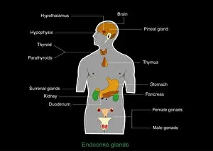
Endocrine system. Computer artwork showing the locations of hormone-producing glands (endocrine glands) in the human body

Background imageSecretory Collection: Exocrine gland types, artwork

Exocrine gland types, artwork
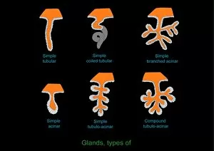
Exocrine gland types. Computer artwork of the six forms an exocrine gland can take. Clockwise from bottom left these are; simple acinar, simple tubular, simple coiled tubular, simple branched acinar

Background imageSecretory Collection: Exocrine gland, artwork

Exocrine gland, artwork
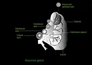
Exocrine gland. Computer artwork of a section through an exocrine gland. Exocrine glands secrete their products through ducts on to epithelial surfaces

Background imageSecretory Collection: Trachea with pollen grains, SEM

Trachea with pollen grains, SEM
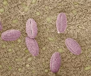
Trachea (windpipe) with pollen grains, coloured scanning electron micrograph (SEM). When airborne particles such as pollen grains are breathed in through the nose or mouth

Background imageSecretory Collection: Small intestine lining, light micrograph

Small intestine lining, light micrograph
Small intestine lining. Light micrograph of a section through the finger-like projections (villi) of the duodenum, the uppermost part of the small intestine

Background imageSecretory Collection: Grape vine, SEM

Grape vine, SEM
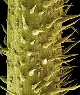
Grape vine. Coloured scanning electron micrograph (SEM) of the surface of a grape vine (Vitis sp.). It is covered in hairs and glandular trichomes (orange)

Background imageSecretory Collection: Sage leaf oil glands, SEM

Sage leaf oil glands, SEM
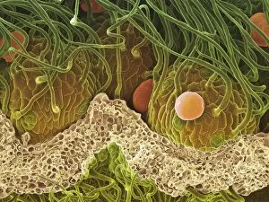
Sage leaf oil glands. Coloured scanning electron micrograph (SEM) of a freeze-fracture through a sage leaf (Salvia officinalis), showing hairs, or trichomes, (green, thin) and oil glands (red)

Background imageSecretory Collection: Thyroid parafollicular cell, TEM

Thyroid parafollicular cell, TEM
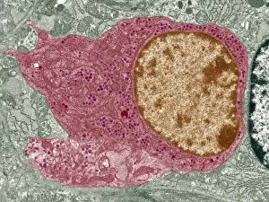
Thyroid parafollicular cell, coloured transmission electron micrograph (TEM). This section has revealed the cells nucleus (light brown)

Background imageSecretory Collection: Breast in pregnancy, light micrograph

Breast in pregnancy, light micrograph
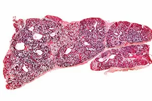
Breast in pregnancy. Light micrograph of a transverse section of the human mammary gland during pregnancy. The breast is normally made up of fibro-fatty connective tissue

Background imageSecretory Collection: Breast tissue, light micrograph

Breast tissue, light micrograph
Breast tissue. Light micrograph of a section through tissue from the human mammary gland during pregnancy. The breast is normally made up of fibro-fatty connective tissue

Background imageSecretory Collection: Skin sweat glands, light micrograph

Skin sweat glands, light micrograph
Skin sweat glands. Light micrograph of a transverse section through skin from a human foot. The skin is made up of an outer epidermis (across top) and a lower epidermis (deep purple)


For sale as Licensed Images

Choose your image, Select your licence and Download the media

"Unveiling the Hidden Secrets: Exploring the Intricacies Cells and Glands" Delving deep into the intricate workings of our body, we uncover a world hints that hold immense significance. From the medulla oblongata in the brain to osteocyte bone cells, each structure plays a vital role in maintaining our well-being. Intriguing artwork showcases the complexity of these secretory systems. The medulla oblongata, often referred to as the "brain's control center, " orchestrates various bodily functions with its network of neurons. Meanwhile, osteocyte bone cells reveal their delicate intricacy under SEM C016 / 9025, highlighting their crucial role in bone formation and maintenance. Moving on to another remarkable organ, we encounter an artistic representation of pancreas anatomy. This gland houses pancreatic islets of Langerhans - clusters of specialized cells responsible for secreting hormones like insulin and glucagon that regulate blood sugar levels. Venturing further into microscopic realms, TEM unveils plasma cells - guardians against infections by producing antibodies that defend our immune system. Thyme leaf oil glands capture attention with their unique secretion properties while contributing to aromatic experiences. SEM images offer glimpses into hidden worlds within us - brain lining displaying its intricate web-like structure; fallopian tube cells showcasing their elegant arrangement; nasal lining revealing its finely textured surface; cervix exhibiting distinctive features under light micrograph F006 / 9805; trachea lining demonstrating its protective capabilities through SEM C013 / 7126. These captivating visualizations remind us of the awe-inspiring complexity present within our bodies' secretory systems. They serve as a testament to nature's ingenuity and inspire us to delve deeper into understanding these hidden secrets that contribute so profoundly to our overall health and well-being.