
Section Collection (#94)
"Exploring the Depths: From Titanic Blueprints to British Submarines, Unveiling Secrets of the Section" Delving into history's archives


For sale as Licensed Images
Choose your image, Select your licence and Download the media
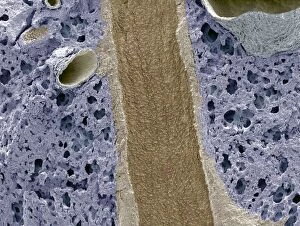
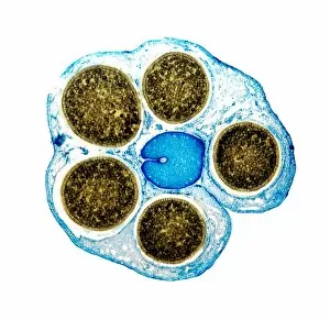
"Exploring the Depths: From Titanic Blueprints to British Submarines, Unveiling Secrets of the Section" Delving into history's archives, we uncover a treasure trove of knowledge through various sections. The intricate blueprint of the Titanic reveals its grandeur and tragic fate, while British submarines spanning five decades shed light on their evolution and contributions to naval warfare. Intriguing our scientific minds, a mesmerizing light micrograph showcases the delicate beauty of cerebellum tissue. Meanwhile, the Higgs boson event captured by ATLAS detector C013/6892 takes us on an exhilarating journey into particle physics. Venturing into architectural wonders, we glimpse St Pauls Cathedral with a section revealing Sir Christopher Wren's genius in designing his iconic dome. A cross-section of Mount Etna unveils its fiery core and reminds us of nature's raw power. Tracing back to wartime struggles, a sectional view exposes the inner workings of a Mills grenade from World War I—a chilling reminder of past conflicts. On a lighter note, Anne Morrow invites us into her imagination as she envisions the interior design of Wates House. The complexity within ourselves is unraveled as we examine a human brain model—an awe-inspiring structure that holds countless mysteries yet to be discovered. Further exploring our anatomy, a diagram guides us through the intricacies of lungs and bronchial tubes—essential for every breath we take. Lastly but not leastly (sic), teeth provide fascinating insights into both adults' and children's dental development—a testament to growth and change throughout life's stages. From engineering marvels like SS Great Britain to artistic interpretations by Anne Morrow—sections offer glimpses beyond what meets the eye; they unravel stories waiting patiently for curious minds to explore their depths.