Sectioned Collection (#45)

"Exploring the Intricacies: From Cerebellum Tissue to Fusion Research, Unveiling the World Sectioned" Delving into the depths of scientific exploration

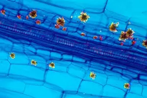
Background imageSectioned Collection: Rhubarb leaf, light micrograph

Rhubarb leaf, light micrograph
Rhubarb leaf. Polarised light micrograph of a longitudinal section through a rhubarb (Rheum sp.) leaf. The dark blue structure is a vascular bundle

Background imageSectioned Collection: Alexander flowers, light micrograph

Alexander flowers, light micrograph
Alexander flowers. Light micrograph of a section through a cluster of alexander flowers (Smyrnium olusatrum). Seen here are cross-sections of the central umbels

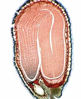
Background imageSectioned Collection: Shepherds purse seed, light micrograph

Shepherds purse seed, light micrograph
Shepherds purse seed. Light micrograph of a longitudinal section through a seed of the shepherds purse (Capsell bursa-pastoris), showing the embryo in the seed

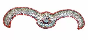
Background imageSectioned Collection: Japanese sago palm leaf, light micrograph

Japanese sago palm leaf, light micrograph
Japanese sago palm leaf. Light micrograph of a transverse section through the leaf (pinna) of a Japanese sago palm, cycad, (Cycas revoluta)

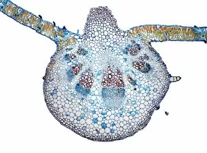
Background imageSectioned Collection: Sunflower leaf, light micrograph

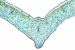
Sunflower leaf, light micrograph
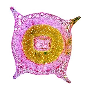
Sunflower leaf. Light micrograph of a transverse section through the midrib of a sunflower (Helianthus annuus) leaf. The upper and lower epidermis on the surfaces of the leaf are blue

Background imageSectioned Collection: Eucalyptus stem, light micrograph

Eucalyptus stem, light micrograph
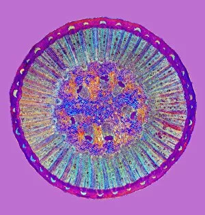
Eucalyptus stem. Light micrograph of a transverse section through a one-year-old stem of a Eucalyptus (Eucalyptus globulus) plant

Background imageSectioned Collection: Mahogany wood structure, light micrograph

Mahogany wood structure, light micrograph
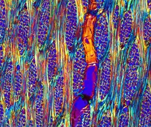
Mahogany wood structure. Polarised light micrograph of a longitudinal tangential section through a woody stem (xylem) of a mahogany (Swietenia sp.) tree

Background imageSectioned Collection: Elm stem, light micrograph

Elm stem, light micrograph
Elm stem (Ulmus procera) section, polarised light micrograph. The black horizontal lines are multiseriate rays, which are composed of several rows of cells

Background imageSectioned Collection: Cucumber flower bud, light micrograph

Cucumber flower bud, light micrograph
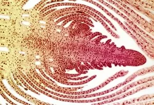
Cucumber flower bud. Light micrograph of a longitudinal section through the flower bud of a cucumber (Cucurbita sp.). The female pistil (red)

Background imageSectioned Collection: Beech leaves, light micrograph

Beech leaves, light micrograph
Beech leaves. Light micrograph of a transverse section through two beech leaves (Fagus sylvatica). The shapes of the two leaves are different because the bottom leaf is constantly exposed to bright
Background imageSectioned Collection: Black pepper stem, light micrograph

Black pepper stem, light micrograph
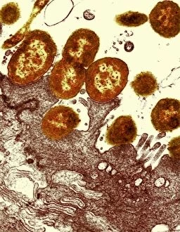
Black pepper stem. Polarised light micrograph of a transverse section through the stem of a black pepper (Pipa nigrum) plant

Background imageSectioned Collection: Poppy fruit, light micrograph

Poppy fruit, light micrograph
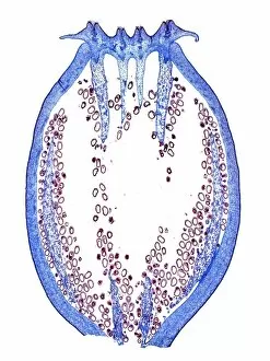
Poppy fruit. Light micrograph of a longitudinal section through the fruit of an Iceland poppy (Papaver nudicaule), showing the seeds (small dots). The fruit capsule has an outer wall (pericarp)

Background imageSectioned Collection: Ginger leaf, light micrograph

Ginger leaf, light micrograph
Ginger leaf. Light micrograph of a transverse section through the midrib of a ginger (Zingiber officinale) leaf. The lower and upper epidermis (blue)

Background imageSectioned Collection: Yew tree leaf, light micrograph

Yew tree leaf, light micrograph
Yew tree leaf. Light micrograph of a transverse section through the leaf (pinna) of a yew tree, (Taxus baccata). The structure has xerophytic (drought plants) characteristics

Background imageSectioned Collection: Birch stem, light micrograph

Birch stem, light micrograph
Birch stem. Light micrograph of a transverse section through the woody stem of a birch (Betula alba) tree, showing a portion of the xylem including some of the pith (bottom left)

Background imageSectioned Collection: Japanese sago palm root, light micrograph

Japanese sago palm root, light micrograph
Japanese sago palm root. Light micrograph of a transverse section through a root of a Japanese sago palm, (Cycas revoluta)

Background imageSectioned Collection: Young yew tree stem, light micrograph

Young yew tree stem, light micrograph
Young yew tree stem. Light micrograph of a transverse section through a one-year-old stem of a yew tree (Taxus baccata). The epidermis (outer layer)

Background imageSectioned Collection: Dandelion root, light micrograph

Dandelion root, light micrograph
Dandelion root. Light micrograph of a transverse section through a root of a dandelion (Taraxacum officinale). The root has undergone secondary thickening

Background imageSectioned Collection: Willow stem, light micrograph

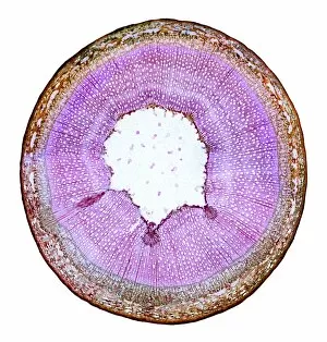
Willow stem, light micrograph
Willow stem. Light micrograph of a cross-section through the stem of a willow (Salix sp.) tree. At centre is the pith (white), surrounded by woody xylem tissue (pink)

Background imageSectioned Collection: Snake intestinal villi, SEM

Snake intestinal villi, SEM
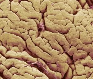
Snake intestine. Coloured scanning electron micrograph (SEM) of villi in the intestine of a snake. The villi (folds) in the surface of the intestine are short and covered in cuboidal mucosal cells

Background imageSectioned Collection: Snake intestine, SEM

Snake intestine, SEM
Snake intestine. Coloured scanning electron micrograph (SEM) of a freeze-fracture through the intestine of a snake. The mucosa (brown)

Background imageSectioned Collection: Reptilian red blood cells, SEM

Reptilian red blood cells, SEM
Reptilian red blood cells. Coloured scanning electron micrograph (SEM) of whole and fractured red blood cells (erythrocytes, purple) from reptilian tissue

Background imageSectioned Collection: Gecko retina, SEM

Gecko retina, SEM
Gecko retina. Coloured scanning electron micrograph (SEM) of an oblique freeze-fracture through the retina of a gecko (family Gekkonidae)

Background imageSectioned Collection: Bone marrow, SEM

Bone marrow, SEM
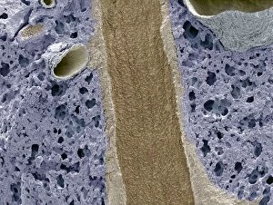
Bone marrow, coloured scanning electron micrograph (SEM). This freeze-fracture has revealed the cavity (lumen) of a large venous sinus (pink), which contains mature blood cells (red)

Background imageSectioned Collection: Wood-boring insect larva

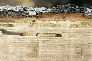
Wood-boring insect larva tunneling through a piece of wood. A generic term for this sort of insect is woodworm. The larvae feed for several years on the interior of wooden objects

Background imageSectioned Collection: Lung bronchus cross-section, SEM

Lung bronchus cross-section, SEM
Lung bronchus cross-section, coloured scanning electron micrograph (SEM). This longitudinal freeze-fracture has exposed the inner epithelial surface of a bronchus (brown)

Background imageSectioned Collection: Salmonella bacterium

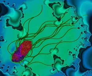
Salmonella bacterium. Coloured transmission electron micrograph (TEM) of a section through a Salmonella sp. bacterium. It is seen on a fractal background

Background imageSectioned Collection: Stenotrophomonas sp. bacteria, TEM

Stenotrophomonas sp. bacteria, TEM
Stenotrophomonas maltophilia bacteria. Coloured transmission electron micrograph (TEM) of a sect- ion through Stenotrophomonas maltophilia bacteria. They are seen on a fractal background

Background imageSectioned Collection: Treponema denticola bacterium, SEM

Treponema denticola bacterium, SEM
Treponema bacterium. Coloured transmission electron micrograph (TEM) of a section through a Treponema denticola bacterium. It is seen on a fractal background

Background imageSectioned Collection: Proteus bacteria

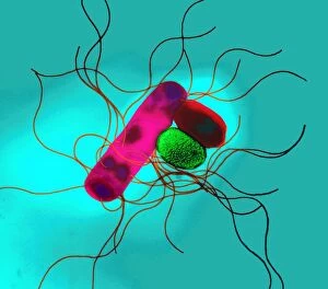
Proteus bacteria. Coloured transmission electron micrograph (TEM) of a section through Proteus sp. bacteria. These Gram-negative bacteria occur in two forms

Background imageSectioned Collection: Campylobacter bacterium, TEM

Campylobacter bacterium, TEM
Campylobacter bacterium. Coloured transmission electron micrograph (TEM) of a section through a Campylobacter sp. bacterium

Background imageSectioned Collection: Helicobacter bacteria, TEM

Helicobacter bacteria, TEM
Helicobacter bacteria. Coloured transmission electron micrograph (TEM) of a section through Helicobacter fennelliae bacteria

Background imageSectioned Collection: Tuberculosis bacteria, TEM

Tuberculosis bacteria, TEM
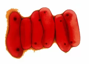
Tuberculosis bacteria. Coloured transmission electron micrograph (TEM) of a section through Mycobacterium tuberculosis bacteria

Background imageSectioned Collection: Capnocytophaga canimorsus bacterium, TEM

Capnocytophaga canimorsus bacterium, TEM
Capnocytophaga canimorsus bacterium. Coloured transmission electron micrograph (TEM) of a sectioned Capnocytophaga canimorsus bacterium

Background imageSectioned Collection: Intestinal bacteria, TEM

Intestinal bacteria, TEM
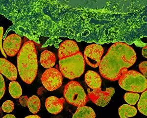
Intestinal bacteria, coloured transmission electron micrograph (TEM). The TEM shows a section through the bacteria (red/yellow) and the wall of the intestines (bottom)

Background imageSectioned Collection: Liverwort spore case, light micrograph

Liverwort spore case, light micrograph
Liverwort spore case. Light micrograph of a vertical section through a spore case (sporophyte) from a liverwort (Pellia epiphylla)

Background imageSectioned Collection: Pine cone scale, light micrograph

Pine cone scale, light micrograph
Pine cone scale. Light micrograph of a vertical section through an ovulate scale from a Scots pine (Pinus sylvestris). This image shows the ovule (oval shape)

Background imageSectioned Collection: Liverwort spore cases, light micrograph

Liverwort spore cases, light micrograph
Liverwort spore cases. Light micrograph of a cross-section through five spore cases (sporophytes) from the liverwort Conocephalum conicum (Marchantiales)

Background imageSectioned Collection: Mycoplasma bacteria, TEM

Mycoplasma bacteria, TEM
Mycoplasma bacteria. Coloured transmission electron micrograph (TEM) of a section through a group of Mycoplasma sp. bacteria (orange) surrounding an infected cell (green)

Background imageSectioned Collection: Maple stem, light micrograph

Maple stem, light micrograph
Maple stem. Polarised light micrograph of a cross-section through the stem of a field maple (Acer campestre). This shows the thick layer of cork (phellem)

Background imageSectioned Collection: Hydrilla bud, light micrograph

Hydrilla bud, light micrograph
Hydrilla bud. Light micrograph of a longitudinal section through an axillary bud on a Hydrilla plant, showing its internal structure

Background imageSectioned Collection: Abnormal thistle stem, light micrograph

Abnormal thistle stem, light micrograph
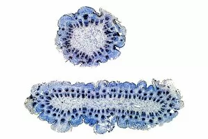
Abnormal thistle stem. Light micrograph of a normal (top) and abnormal (bottom) stem of the perennial sow thistle (Sonchus arvensis). The abnormal stem has grown in a flattened, or fasciated, manner

Background imageSectioned Collection: Nettle leaf stalk structures, LM

Nettle leaf stalk structures, LM
Nettle leaf stalk structures. Polarised light micrograph (LM) of a nettle leaf (Urtica dioica), which has been boiled up with dilute sodium hydroxide to separate the cells

Background imageSectioned Collection: Water-lily leaf stem, light micrograph

Water-lily leaf stem, light micrograph
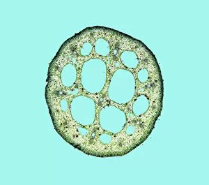
Water-lily leaf stem. Light micrograph of a cross- section through the leaf stem (petiole) of a white water-lily (Nymphaea alba)
Background imageSectioned Collection: Stem section, SEM

Stem section, SEM
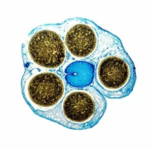
Stem section. Coloured scanning electron micrograph (SEM) of a section through a stem of a woody dicotyledon plant. This stem consists mainly of secondary xylem tissue (orange, centre)

Background imageSectioned Collection: Walnut bark

Walnut bark. Cross section through the trunk of a walnut tree (Juglans regia) revealing the pattern in its bark. This pattern is known as a rhytidome

Background imageSectioned Collection: Sectioned leaf, SEM

Sectioned leaf, SEM
Leaf tissue structure. Coloured scanning electron micrograph (SEM) of a freeze-fracture through a rose plant (Rosa sp. ) leaf. The leaf cells are the rectangular shapes in the fracture plane

Background imageSectioned Collection: Bamboo stem, SEM

Bamboo stem, SEM
Bamboo stem. Coloured scanning electron micrograph (SEM) of a section through a bamboo stem (Phyllostachys sp.). Vascular bundles can be seen (bright yellow) containing xylem (larger openings)


For sale as Licensed Images

Choose your image, Select your licence and Download the media

"Exploring the Intricacies: From Cerebellum Tissue to Fusion Research, Unveiling the World Sectioned" Delving into the depths of scientific exploration, a light micrograph reveals the intricate structure of cerebellum tissue. Witnessing a Higgs boson event through the lens of ATLAS detector C013/6892, scientists capture an awe-inspiring moment in particle physics. A meticulously crafted model showcases the complexity and beauty of the human brain, inviting us to marvel at its wonders. Peering into hippocampus brain tissue under a microscope, we uncover secrets that may hold keys to memory formation and learning processes. In pursuit of limitless energy sources, fusion research takes center stage as we examine a tokamak device designed for controlled nuclear fusion reactions. The inner workings of our brains come alive through an MRI scan, revealing detailed brain anatomy like never before imagined. Illuminated by light microscopy, dicotyledon plant stem sections unveil nature's architectural masterpiece in stunning detail. EDTA crystals take on mesmerizing patterns when viewed under intense magnification - showcasing their unique molecular arrangement through light micrography. Journeying deep within our bodies, kidney tubules are sectioned to shed light on their vital role in maintaining fluid balance and waste removal systems. An X-ray image exposes every contour and intricacy of a human skull – offering glimpses into our skeletal framework with remarkable clarity. Through transmission electron microscopy (TEM), rough endoplasmic reticulum emerges as an interconnected network responsible for protein synthesis within cells' cytoplasmic matrix Computer artwork visualizes Alzheimer's-affected brain regions – reminding us why continued research is crucial in unraveling this devastating disease. In this captivating journey across various scientific disciplines and microscopic landscapes, "sectioned" unveils hidden worlds that expand our understanding of the universe and ourselves.