Sectioned Collection (#49)

"Exploring the Intricacies: From Cerebellum Tissue to Fusion Research, Unveiling the World Sectioned" Delving into the depths of scientific exploration

Background imageSectioned Collection: Sugar cube crystals, SEM

Sugar cube crystals, SEM
Sugar cube crystals. Coloured scanning electron micrograph (SEM) of a freeze-fracture through a sugar cube. The fracture plane shows the sucrose crystals that make up the sugar cube

Background imageSectioned Collection: Dogfish head, transverse section

Dogfish head, transverse section
Dogfish head. Light micrograph of a transverse section through the head of a lesser spotted dogfish (Scyliorhinus canicula), in the region of the eyes (upper left and upper right)

Background imageSectioned Collection: Intervertebral disc, light micrograph

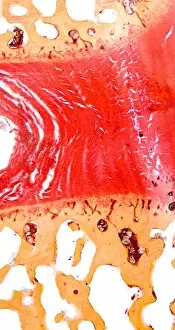
Intervertebral disc, light micrograph
Intervertebral disc. Light micrograph of a transverse section through an intervertebral disc (red) and the surrounding tissue (brown)

Background imageSectioned Collection: Heel skin tissue, light micrograph

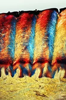
Heel skin tissue, light micrograph
Heel skin tissue. Polarised light micrograph of a transverse section through skin from the heel of a human foot. The sole of the foot has to withstand the weight of the body

Background imageSectioned Collection: Lamprey gill region, light micrograph

Lamprey gill region, light micrograph
Lamprey gill. Light micrograph of a transverse section through the gill region of a lamprey (Petromyzon marinus), a parasitic fish. The spinal area is at upper centre

Background imageSectioned Collection: Breast tissue, light micrograph

Breast tissue, light micrograph
Breast tissue. Light micrograph of a section through tissue from the human mammary gland during pregnancy. The breast is normally made up of fibro-fatty connective tissue

Background imageSectioned Collection: Rabbit eye, longitudinal section

Rabbit eye, longitudinal section
Rabbit eye. Light micrograph of a longitudinal section through the eye of a rabbit (Oryctolagus cuniculus). The rabbit has a typical mammalian eye structure

Background imageSectioned Collection: Plasma cell, TEM

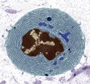
Plasma cell, TEM
Plasma cell, coloured transmission electron micrograph (TEM). This section has revealed the cells large central nucleus (brown)

Background imageSectioned Collection: Shark skin, light micrograph

Shark skin, light micrograph
Shark skin. Light micrograph of a section through the skin of a shark, showing two dermal denticles. These bony structures cover the whole skin of the shark

Background imageSectioned Collection: Taste buds, light micrograph

Taste buds, light micrograph
Taste buds. Coloured light micrograph of a section through the tongue, showing taste buds (round, purple). The taste buds are within papillae (projections) located on the surface of the tongue

Background imageSectioned Collection: Stomach pylorus glands, light micrograph

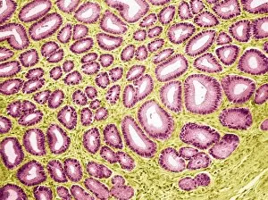
Stomach pylorus glands, light micrograph
Stomach pylorus glands. Light micrograph of a section through glands (pink) in the pylorus region of the stomach. Pyloric glands contain mucus cells and G cells that secrete gastrin

Background imageSectioned Collection: Thyroid follicle structures, TEM

Thyroid follicle structures, TEM
Thyroid follicle structures. Coloured transmission electron micrograph (TEM) of a section through structures in a thyroid follicle

Background imageSectioned Collection: Sheeps hoof, light micrograph

Sheeps hoof, light micrograph
Sheeps hoof. Polarised light micrograph of a transverse section through the hoof of a sheep. In sheep and other hooved animals, the hoof is made of the same material as nails and claws

Background imageSectioned Collection: Pneumonia infection, light micrograph

Pneumonia infection, light micrograph
Pneumonia infection. Light micrograph of a section through lung tissue from a patient with bronchial pneumonia. Shown here is the blockage and breakdown of the air sacs

Background imageSectioned Collection: Sago cycad leaf, SEM

Sago cycad leaf, SEM
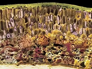
Sago cycad leaf. Coloured scanning electron micrograph (SEM) of a section through a sago cycad (Cycas revoluta) leaf. At lower centre and left are vascular bundles (orange clusters)

Background imageSectioned Collection: Chlamydomonas alga

Chlamydomonas alga
Coloured Transmission Electron Micrograph of a section through the unicellular green algae, Chlamydomonas asymmetrica. The micrograph shows the arrangement of chloroplasts (green)

Background imageSectioned Collection: Mushroom, light micrograph

Mushroom, light micrograph
Mushroom. Light micrograph (LM) of a section through a mushroom, Agaricus sp. (formerly Psalliota sp.), showing its stalk (orange) and gills (long white projections)

Background imageSectioned Collection: Mushroom gills, light micrograph

Mushroom gills, light micrograph
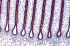
Mushroom gills. Light micrograph (LM) of a section through the gills of a mushroom, Agaricus sp. (formerly Psalliota sp.)

Background imageSectioned Collection: Spinal cord, transverse section

Spinal cord, transverse section
Spinal cord. Light micrograph of a transverse section through a human spinal cord. The spinal cord has a central canal around which are the dorsal and ventral horns (orange)

Background imageSectioned Collection: Dog tongue tissue, light micrograph

Dog tongue tissue, light micrograph
Dog tongue tissue. Light micrograph of a transverse section through the tongue of a dog, showing the tongues surface (across top)

Background imageSectioned Collection: Alzheimers disease, artwork

Alzheimers disease, artwork
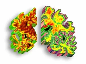
Alzheimers disease. Computer artwork of a vertical (coronal) slice through the brain of an Alzheimer patient (left) compared with a normal brain (right)

Background imageSectioned Collection: Foot skin tissue, light micrograph

Foot skin tissue, light micrograph
Foot skin tissue. Light micrograph of a transverse section through skin from a human foot. The skin is made up of an outer epidermis (across top)

Background imageSectioned Collection: Brain tissue, light micrograph

Brain tissue, light micrograph
Brain tissue. Light micrograph of a section through tissue from the mammalian brain, in this case from the cerebellum. The cerebellum is the part of the brain that controls sensory perception

Background imageSectioned Collection: Skin sweat glands, light micrograph

Skin sweat glands, light micrograph
Skin sweat glands. Light micrograph of a transverse section through skin from a human foot. The skin is made up of an outer epidermis (across top) and a lower epidermis (deep purple)

Background imageSectioned Collection: Brain complexity, conceptual artwork

Brain complexity, conceptual artwork
Brain complexity. Conceptual computer artwork of a slice through a brain represented as a complex maze, with a question mark at its centre

Background imageSectioned Collection: Cat tongue tissue, light micrograph

Cat tongue tissue, light micrograph
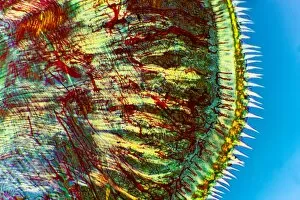
Cat tongue tissue. Polarised light micrograph of a transverse section through a cats tongue. The outer stratified epithelium (right) has spike-like (white) keratinised papillae

Background imageSectioned Collection: Tongue tissue, light micrograph

Tongue tissue, light micrograph
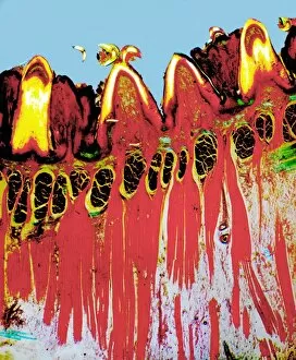
Tongue tissue. Polarised light micrograph of a transverse section through a tongue, showing the surface (across top). Here, there are four rounded fungiform papillae

Background imageSectioned Collection: Experiments with Fairlies steam carriage

Experiments with Fairlies steam carriage
Fairlies steam carriage; an invention that gained much criticism, due to its unusual seating design. The train is sectioned into 2 first class carriages seating 16 people

Background imageSectioned Collection: Bacterial cell

Bacterial cell, computer artwork. This is ageneralised bacterium, illustrating features ofdifferent bacterial types that would not becombined in a single cell


For sale as Licensed Images

Choose your image, Select your licence and Download the media

"Exploring the Intricacies: From Cerebellum Tissue to Fusion Research, Unveiling the World Sectioned" Delving into the depths of scientific exploration, a light micrograph reveals the intricate structure of cerebellum tissue. Witnessing a Higgs boson event through the lens of ATLAS detector C013/6892, scientists capture an awe-inspiring moment in particle physics. A meticulously crafted model showcases the complexity and beauty of the human brain, inviting us to marvel at its wonders. Peering into hippocampus brain tissue under a microscope, we uncover secrets that may hold keys to memory formation and learning processes. In pursuit of limitless energy sources, fusion research takes center stage as we examine a tokamak device designed for controlled nuclear fusion reactions. The inner workings of our brains come alive through an MRI scan, revealing detailed brain anatomy like never before imagined. Illuminated by light microscopy, dicotyledon plant stem sections unveil nature's architectural masterpiece in stunning detail. EDTA crystals take on mesmerizing patterns when viewed under intense magnification - showcasing their unique molecular arrangement through light micrography. Journeying deep within our bodies, kidney tubules are sectioned to shed light on their vital role in maintaining fluid balance and waste removal systems. An X-ray image exposes every contour and intricacy of a human skull – offering glimpses into our skeletal framework with remarkable clarity. Through transmission electron microscopy (TEM), rough endoplasmic reticulum emerges as an interconnected network responsible for protein synthesis within cells' cytoplasmic matrix Computer artwork visualizes Alzheimer's-affected brain regions – reminding us why continued research is crucial in unraveling this devastating disease. In this captivating journey across various scientific disciplines and microscopic landscapes, "sectioned" unveils hidden worlds that expand our understanding of the universe and ourselves.