
Spacefilled Collection (#4)
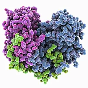
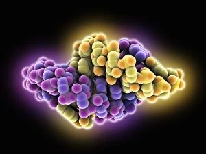
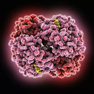
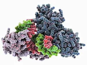
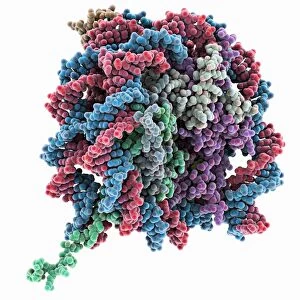
"Exploring the Vastness of Spacefilled: From Molecules to Medicines" Delving into the intricate world of molecules, we encounter the Cytochrome b5 molecule C015 / 6696


For sale as Licensed Images
Choose your image, Select your licence and Download the media
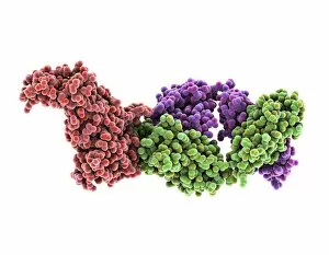
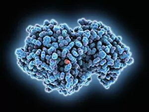
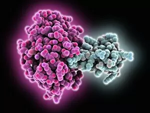
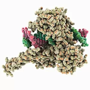
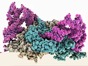
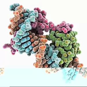
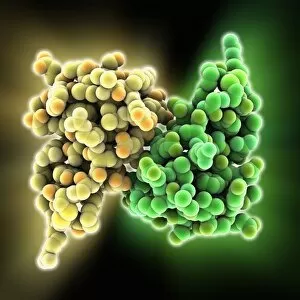
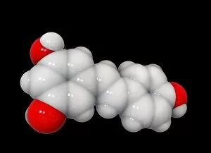
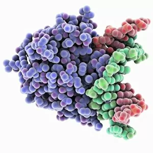
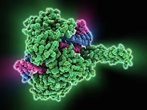
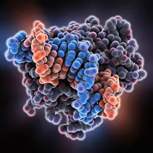
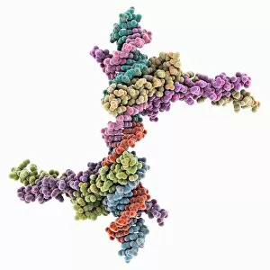
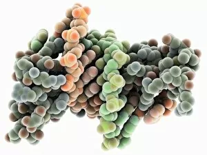
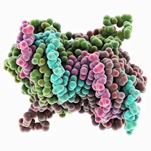
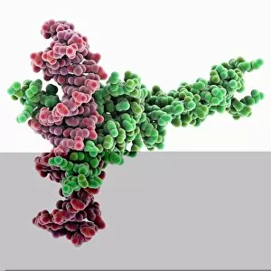
"Exploring the Vastness of Spacefilled: From Molecules to Medicines" Delving into the intricate world of molecules, we encounter the Cytochrome b5 molecule C015 / 6696, a key player in electron transfer processes within cells. Unraveling the mysteries of DNA structure, we come across the Z-DNA tetramer molecule C015 / 6557, showcasing its unique left-handed helical conformation. Embarking on a psychedelic journey, we discover the Psilocybin drug molecule, known for its mind-altering effects and potential therapeutic applications. Witnessing precision at work, we examine the RNA-editing enzyme molecular model that plays a crucial role in modifying genetic information to ensure cellular functionality. Energizing our bodies from within, we explore the ATPase molecule responsible for powering various cellular processes by converting ATP into ADP and phosphate. Battling against viral invaders, we observe Rhinovirus and antibody molecular models (C015 / 7139 & C015 / 7138) engaged in an intricate dance of recognition and defense. Admiring art imitating life, we marvel at an artwork depicting DNA's elegant double helix structure (C017 / 7217), symbolizing life's blueprint encoded within our genes. Nourishing our bodies with essential building blocks, Valine molecule takes center stage as one of the amino acids vital for protein synthesis and muscle repair. Embracing metabolic pathways with Methionine molecule as our guide; this sulfur-containing amino acid is indispensable for protein synthesis and methylation reactions. Discovering Histidine's multifaceted nature - not only serving as an amino acid but also playing pivotal roles in pH regulation and metal ion coordination within proteins.