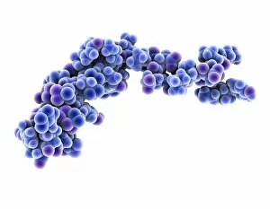
Spacefilled Collection (#7)

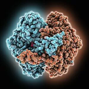
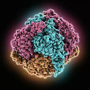
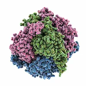
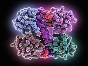
"Exploring the Vastness of Spacefilled: From Molecules to Medicines" Delving into the intricate world of molecules, we encounter the Cytochrome b5 molecule C015 / 6696

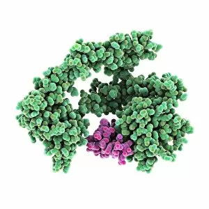
Background imageSpacefilled Collection: Hepatitis D virus ribozyme complex

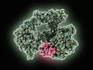
Hepatitis D virus ribozyme complex. Computer model showing an RNA (ribonucleic acid) strand from an Hepatitis delta (Hepatitis D) virus genomic ribozyme, complexed with a ribonucleoprotein

Background imageSpacefilled Collection: Lumazine synthase molecule

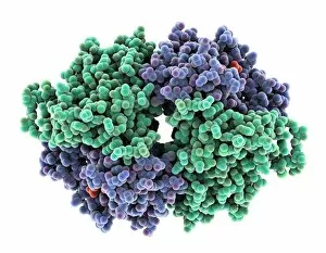
Lumazine synthase molecule. Computer model showing the structure of a lumazine synthase enzyme molecule from a Brucella abortus bacterium

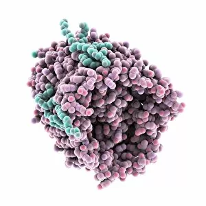
Background imageSpacefilled Collection: Poly(A)-binding protein and RNA complex

Poly(A)-binding protein and RNA complex. Computer model showing the structure of a poly(A)-binding protein (PABP) molecule bound to the poly(A)

Background imageSpacefilled Collection: Vitamin B1 molecule

Vitamin B1 molecule. Computer model showing the structure of a molecule of vitamin B1 (thiamine). Vitamin B1 is an essential nutrient that humans are unable to produce

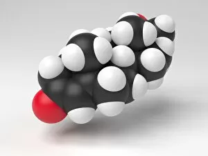
Background imageSpacefilled Collection: Testosterone hormone molecule

Testosterone hormone molecule. Computer model showing the structure of a molecule of the male sex hormone testosterone. Testosterone is the main human androgen

Background imageSpacefilled Collection: Progesterone hormone molecule

Progesterone hormone molecule. Computer model showing the structure of a molecule of the hormone progesterone. Progesterone is produced in the ovaries of women and the testes of men

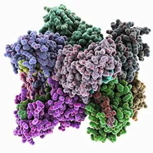
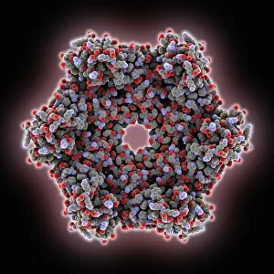
Background imageSpacefilled Collection: ATPase molecule C015 / 7073

ATPase molecule C015 / 7073
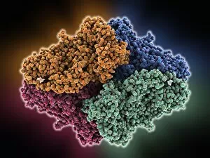
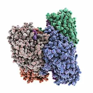
ATPase molecule. Molecular model of the ATP-dependent oligomerisation domain of N-ethylmaleimide sensitive factor (NSF), a hexameric ATPase

Background imageSpacefilled Collection: Thrombin protein, molecular model C015 / 7074

Thrombin protein, molecular model C015 / 7074
Thrombin protein, molecular model. Thrombin is an enzyme involved in the blood coagulation (clotting) process. It converts fibrinogen (a soluble plasma glycoprotein synthesised in the liver)

Background imageSpacefilled Collection: Caffeine molecule

Caffeine molecule. Computer artwork showing the structure of a molecule of the alkaloid stimulant and legal drug caffeine. Caffeine is found in drinks such as tea, coffee, and fizzy drinks

Background imageSpacefilled Collection: Phosphoglucose isomerase molecule C015 / 7036

Phosphoglucose isomerase molecule C015 / 7036
Phosphoglucose isomerase molecule. Molecular model of a rabbit phosphoglucose isomerase molecule complexed with a fructose-6-phosphate molecule

Background imageSpacefilled Collection: Flavocytochrome b2 molecule C015 / 7033

Flavocytochrome b2 molecule C015 / 7033
Flavocytochrome b2, molecular model. This enzyme is found in the intermembrane space of mitochondria, where it catalyses the oxidation of lactate to pyruvate

Background imageSpacefilled Collection: Restriction enzyme and DNA C015 / 6941

Restriction enzyme and DNA C015 / 6941
Restriction enzyme and DNA. Molecular model showing an EcoRI endonuclease enzyme (purple) bound to a DNA (deoxyribonucleic acid) strand (blue). EcoRI is an enzyme isolated from strains of E

Background imageSpacefilled Collection: Human serum albumin molecule C015 / 6938

Human serum albumin molecule C015 / 6938
Human serum albumin, molecular model. Albumin is the most abundant protein in human blood plasma. It is seen here complexed with stearic acid, a saturated fatty acid

Background imageSpacefilled Collection: Dienoyl-coenzyme A isomerase molecule C015 / 6852

Dienoyl-coenzyme A isomerase molecule C015 / 6852
Dienoyl-coenzyme A isomerase molecule. Computer model showing the structure of a molecule of dienoyl-coenzyme A (dienoyl-CoA) isomerase

Background imageSpacefilled Collection: Dienoyl-coenzyme A isomerase molecule C015 / 6827

Dienoyl-coenzyme A isomerase molecule C015 / 6827
Dienoyl-coenzyme A isomerase molecule. Computer model showing the structure of a molecule of dienoyl-coenzyme A (dienoyl-CoA) isomerase

Background imageSpacefilled Collection: Z-Z DNA junction, molecular model C016 / 1539

Z-Z DNA junction, molecular model C016 / 1539
Z-Z DNA junction. Molecular model of a junction between two left-handed DNA double helices (Z DNA, down centre) stabilised by the Z alpha domain of the human RNA-editing enzyme ADAR1

Background imageSpacefilled Collection: Cytotoxic T-lymphocyte antigen-4 C016 / 1633

Cytotoxic T-lymphocyte antigen-4 C016 / 1633
Cytotoxic T-lymphocyte antigen-4 (CTLA-4), molecular model. The protein receptor is found on T-lymphocytes, one of the cells of the immune system

Background imageSpacefilled Collection: Cytotoxic T-lymphocyte antigen-4 C016 / 1567

Cytotoxic T-lymphocyte antigen-4 C016 / 1567
Cytotoxic T-lymphocyte antigen-4 (CTLA-4), molecular model. The protein receptor is found on T-lymphocytes, one of the cells of the immune system

Background imageSpacefilled Collection: Z-Z DNA junction, molecular model C016 / 1445

Z-Z DNA junction, molecular model C016 / 1445
Z-Z DNA junction. Molecular model of a junction between two left-handed DNA double helices (Z DNA, pink) stabilised by the Z alpha domain of the human RNA-editing enzyme ADAR1

Background imageSpacefilled Collection: Beta-galactosidase molecule C015 / 1971

Beta-galactosidase molecule C015 / 1971
Beta-galactosidase, molecular model. This enzyme breaks down sugars containing galactose, such as lactose, into their basic units (monosaccharides)

Background imageSpacefilled Collection: Beta-galactosidase molecule C015 / 1970

Beta-galactosidase molecule C015 / 1970
Beta-galactosidase, molecular model. This enzyme breaks down sugars containing galactose, such as lactose, into their basic units (monosaccharides)

Background imageSpacefilled Collection: Cytochrome b5 molecule C015 / 6695

Cytochrome b5 molecule C015 / 6695
Cytochrome b5. Molecular model of cytochrome b5 from a cows liver. Cytochrome molecules perform oxidation and reduction reactions for electron transport

Background imageSpacefilled Collection: Citrate synthase molecule C015 / 6694

Citrate synthase molecule C015 / 6694
Citrate synthase, molecular model. This enzyme is involved in the first step of the citric acid (or Krebs) cycle, the process by which mitochondria convert glucose to energy

Background imageSpacefilled Collection: Citrate synthase molecule C015 / 6693

Citrate synthase molecule C015 / 6693
Citrate synthase, molecular model. This enzyme is involved in the first step of the citric acid (or Krebs) cycle, the process by which mitochondria convert glucose to energy

Background imageSpacefilled Collection: Type IV collagen C4 domain molecule C015 / 5574

Type IV collagen C4 domain molecule C015 / 5574
Type IV collagen C4 domain. Molecular model of the type IV collagen C4 domain found at the C-terminus of type IV collagens

Background imageSpacefilled Collection: Type IV collagen C4 domain molecule C015 / 5573

Type IV collagen C4 domain molecule C015 / 5573
Type IV collagen C4 domain. Molecular model of the type IV collagen C4 domain found at the C-terminus of type IV collagens

Background imageSpacefilled Collection: Influenza A non-structural-protein 1 C015 / 5927

Influenza A non-structural-protein 1 C015 / 5927
Influenza A non-structural-protein 1 (ns1), molecular model. This RNA-binding protein is needed for viral replication. It binds to host mRNA (messenger ribonucleic acid)

Background imageSpacefilled Collection: Influenza A non-structural protein 1 C015 / 5926

Influenza A non-structural protein 1 C015 / 5926
Influenza A non-structural protein 1 (ns1), molecular model. This RNA-binding protein is needed for viral replication. It binds to host mRNA (messenger ribonucleic acid)

Background imageSpacefilled Collection: Epidermal growth factor molecule C015 / 5925

Epidermal growth factor molecule C015 / 5925
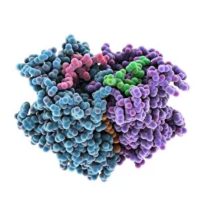
Epidermal growth factor. Molecular model of human epidermal growth factor (EGF, green) complexed with its receptor (pink)

Background imageSpacefilled Collection: Epidermal growth factor molecule C015 / 5924

Epidermal growth factor molecule C015 / 5924
Epidermal growth factor. Molecular model of human epidermal growth factor (EGF, green) complexed with its receptor (pink)

Background imageSpacefilled Collection: Z-DNA tetramer molecule C015 / 6558

Z-DNA tetramer molecule C015 / 6558
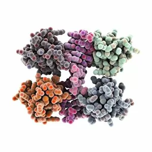
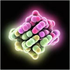
Z-DNA (deoxyribonucleic acid) tetramer, molecular model. DNA is composed of two strands twisted into a double helix. This is a tetramer of the molecule, containing four strands

Background imageSpacefilled Collection: Monoclonal antibody fragment C015 / 6556

Monoclonal antibody fragment C015 / 6556
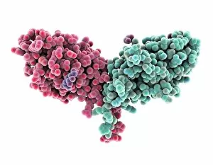
Monoclonal antibody fragment. Molecular model of the antigen-binding fragment (Fab) of the monoclonal antibody cetuximab. Cetuximab is a chemotherapy drug, used to treat metastatic colon cancer

Background imageSpacefilled Collection: Monoclonal antibody fragment C015 / 6555

Monoclonal antibody fragment C015 / 6555
Monoclonal antibody fragment. Molecular model of the antigen-binding fragment (Fab) of the monoclonal antibody cetuximab. Cetuximab is a chemotherapy drug, used to treat metastatic colon cancer

Background imageSpacefilled Collection: Vitamin B12 transport protein C015 / 5824

Vitamin B12 transport protein C015 / 5824
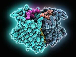
Vitamin B12 transport protein, molecular model. This transmembrane protein, known as BTUB, is from the Escherichia coli bacterium

Background imageSpacefilled Collection: Vitamin B12 transport protein C015 / 5823

Vitamin B12 transport protein C015 / 5823
Vitamin B12 transport protein, molecular model. This transmembrane protein, known as BTUB, is from the Escherichia coli bacterium

Background imageSpacefilled Collection: HIV-1 polypurine tract, molecular model C015 / 5821

HIV-1 polypurine tract, molecular model C015 / 5821
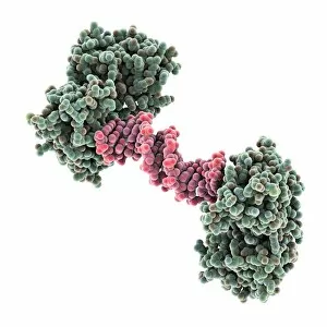
HIV-1 polypurine tract. Molecular model of the polypurine tract (PPT, red) from human immunodeficiency virus-1 (HIV-1) complexed with reverse transcriptase (green)

Background imageSpacefilled Collection: Androgen receptor, molecular model C015 / 6113

Androgen receptor, molecular model C015 / 6113
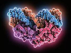
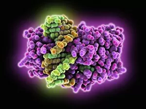
Androgen receptor. Molecular model of the DNA-binding region of an androgen receptor (purple and red) complexed with DNA (deoxyribonucleic acid, blue and orange)

Background imageSpacefilled Collection: Outer membrane phospholipase A molecule C015 / 6111

Outer membrane phospholipase A molecule C015 / 6111
Outer membrane phospholipase A. Molecular model of the integral membrane protein, outer membrane phospholipase A from the Escherichia coli bacterium

Background imageSpacefilled Collection: Androgen receptor, molecular model C015 / 6112

Androgen receptor, molecular model C015 / 6112
Androgen receptor. Molecular model of the DNA-binding region of an androgen receptor (purple and brown) complexed with DNA (deoxyribonucleic acid, turquoise and red)

Background imageSpacefilled Collection: Outer membrane phospholipase A molecule C015 / 6110

Outer membrane phospholipase A molecule C015 / 6110
Outer membrane phospholipase A. Molecular model of the integral membrane protein, outer membrane phospholipase A from the Escherichia coli bacterium

Background imageSpacefilled Collection: Isovaleryl dehydrogenase enzyme C015 / 5439

Isovaleryl dehydrogenase enzyme C015 / 5439
Isovaleryl dehydrogenase. Molecular model of the human isovaleryl-CoA dehydrogenase (IVD) enzyme. IVD is an oxidoreductase enzyme that catalyses the third step in the break down of the amino acid

Background imageSpacefilled Collection: ATPase molecule

ATPase molecule. Computer model showing the structure of the peripheral stalk (stator) of an ATP synthase (ATPase) molecule from a cow mitochondrion

Background imageSpacefilled Collection: Isovaleryl dehydrogenase enzyme C015 / 5438

Isovaleryl dehydrogenase enzyme C015 / 5438
Isovaleryl dehydrogenase. Molecular model of the human isovaleryl-CoA dehydrogenase (IVD) enzyme. IVD is an oxidoreductase enzyme that catalyses the third step in the break down of the amino acid

Background imageSpacefilled Collection: Methyltransferase and DNA C015 / 5704

Methyltransferase and DNA C015 / 5704
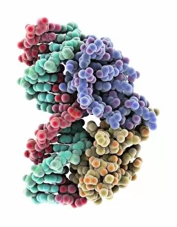
Methyltransferase and DNA. Molecular model of the enzyme HhaI methyltransferase (purple) complexed with a molecule of DNA (deoxyribonucleic acid, orange and green)

Background imageSpacefilled Collection: Resolvase complexed with DNA

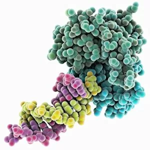
Resolvase complexed with DNA. Molecular model of RusA resolvase (blue and green) bound to a DNA (deoxyribonucleic acid, pink and yellow) duplex

Background imageSpacefilled Collection: HGPRTase molecule C015 / 5405

HGPRTase molecule C015 / 5405
HGPRTase. Molecular model of hypoxanthine-guanine phosphoribosyltransferase (HGPRTase) bound to a molecule of guanosine monophosphate (GMP). HGPRTase is involved in the purine salvage pathway

Background imageSpacefilled Collection: Methyltransferase and DNA C015 / 5703

Methyltransferase and DNA C015 / 5703
Methyltransferase and DNA. Molecular model of the enzyme HhaI methyltransferase (purple) complexed with a molecule of DNA (deoxyribonucleic acid, yellow and green)

Background imageSpacefilled Collection: Neuropeptide Y neurotransmitter molecule C015 / 6240

Neuropeptide Y neurotransmitter molecule C015 / 6240
Neuropeptide Y neurotransmitter molecule. Molecular model showing the structure of the neurotransmitter neuropeptide Y (NPY). NPY is found in the brain and autonomic nervous system


For sale as Licensed Images

Choose your image, Select your licence and Download the media

"Exploring the Vastness of Spacefilled: From Molecules to Medicines" Delving into the intricate world of molecules, we encounter the Cytochrome b5 molecule C015 / 6696, a key player in electron transfer processes within cells. Unraveling the mysteries of DNA structure, we come across the Z-DNA tetramer molecule C015 / 6557, showcasing its unique left-handed helical conformation. Embarking on a psychedelic journey, we discover the Psilocybin drug molecule, known for its mind-altering effects and potential therapeutic applications. Witnessing precision at work, we examine the RNA-editing enzyme molecular model that plays a crucial role in modifying genetic information to ensure cellular functionality. Energizing our bodies from within, we explore the ATPase molecule responsible for powering various cellular processes by converting ATP into ADP and phosphate. Battling against viral invaders, we observe Rhinovirus and antibody molecular models (C015 / 7139 & C015 / 7138) engaged in an intricate dance of recognition and defense. Admiring art imitating life, we marvel at an artwork depicting DNA's elegant double helix structure (C017 / 7217), symbolizing life's blueprint encoded within our genes. Nourishing our bodies with essential building blocks, Valine molecule takes center stage as one of the amino acids vital for protein synthesis and muscle repair. Embracing metabolic pathways with Methionine molecule as our guide; this sulfur-containing amino acid is indispensable for protein synthesis and methylation reactions. Discovering Histidine's multifaceted nature - not only serving as an amino acid but also playing pivotal roles in pH regulation and metal ion coordination within proteins.