Spore Collection (#5)

"Exploring the Fascinating World of Spores: From Aspergillus to Fungal Kingdom" Delving into the microscopic realm, we uncover the enchanting world of spores

Background imageSpore Collection: Wax Cap Fungus - UK

Wax Cap Fungus - UK
DAC-613 Wax Cap Fungus UK Hygrocybe ceracea David Chapman Please note that prints are for personal display purposes only and may not be reproduced in any way

Background imageSpore Collection: Lycopod

Lycopod

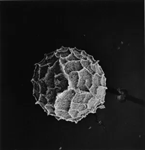
Background imageSpore Collection: Selaginella kraussiana, spikemoss

Selaginella kraussiana, spikemoss
Scanning electron microscope image of the female spore of Krauss spikemoss (x 150 on a standard 9 cm wide print)

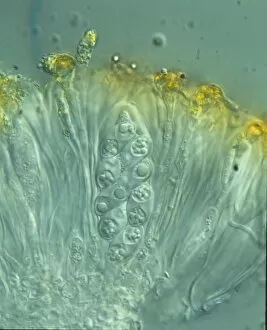
Background imageSpore Collection: Xanthoria parietina, lichen

Xanthoria parietina, lichen
Shown here is a maritime sunburst lichen. A photograph of the ascus containing eight ascopores

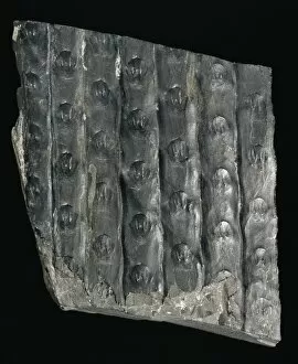
Background imageSpore Collection: Sigillaria laevigata, fossil lycopod bark

Sigillaria laevigata, fossil lycopod bark
This carboniferous fossilised lycopod (prehistoric spore-bearing plants) bark specimen is 9 cm long

Background imageSpore Collection: Cystopteris diaphana, diaphanous bladder fern

Cystopteris diaphana, diaphanous bladder fern
An SEM showing a close-up of the spiny-lacunar surface of the diaphanous bladder fern (Cystopteris diaphana) spore. Photographed using Philips XL30 SEM

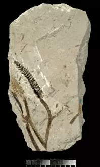
Background imageSpore Collection: Protobarinophyton obrutschevii

Protobarinophyton obrutschevii
The fossilised leafless stems of Protobarinophyton obrutschevii topped by a cone-like array of sopre sacs. Approximately 3 mm wide

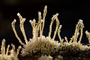
Background imageSpore Collection: Wall Screw-moss (Tortula muralis) frost covered spore capsules, growing on concrete wall, Powys

Wall Screw-moss (Tortula muralis) frost covered spore capsules, growing on concrete wall, Powys, Wales, January

Background imageSpore Collection: Oak Bonnet-cap - tree trunk - UK

Oak Bonnet-cap - tree trunk - UK
DAC-94 Oak Bonnet-cap - tree trunk UK Mycena inclinata David Chapman Please note that prints are for personal display purposes only and may not be reproduced in anyway

Background imageSpore Collection: Sword Fern, (Polystichum munitum), spores, Cascade Mountains, Washington, USA

Sword Fern, (Polystichum munitum), spores, Cascade Mountains, Washington, USA

Background imageSpore Collection: Bootlaces of Honey Fungus

Bootlaces of Honey Fungus
DAC-4 Bootlaces of Honey Fungus Armillaria mellea David Chapman contact details: prints@ardea.com tel: +44 (0) 20 8318 1401

Background imageSpore Collection: Common Earthball - Godolphin Woods - Cornwall - UK

Common Earthball - Godolphin Woods - Cornwall - UK
DAC-84 Common Earthball - Godolphin Woods Cornwall - UK Scleroderma citrinum David Chapman contact details: prints@ardea.com tel: +44 (0) 20 8318 1401

Background imageSpore Collection: Earth Star - shedding spores - Bedfordshire UK 14918

Earth Star - shedding spores - Bedfordshire UK 14918
BB-2632 Earth Star - shedding spores Bedfordshire UK Geastrum sp Brian Bevan contact details: prints@ardea.com tel: +44 (0) 20 8318 1401

Background imageSpore Collection: Orange Peel Fungus - Cornwall - UK

Orange Peel Fungus - Cornwall - UK
DAC-285 Orange Peel Fungus Cornwall - UK Aleuria aurantia David Chapman contact details: prints@ardea.com tel: +44 (0) 20 8318 1401

Background imageSpore Collection: Many Zoned Polypore

Many Zoned Polypore
DAC-31 Many Zoned Polypore Treametes versicolor David Chapman contact details: prints@ardea.com tel: +44 (0) 20 8318 1401

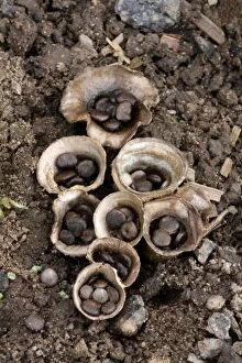
Background imageSpore Collection: Field Birds Nest Fungus (Crucibulum laeve) fruiting bodies, splash cups with peridiole spore

Field Birds Nest Fungus (Crucibulum laeve) fruiting bodies, splash cups with peridiole spore capsules, adapted for spore dispersal by raindrops, growing in agricultural land, Leicestershire, England

Background imageSpore Collection: Dance Fly (Empis tessellata) adult, fallen victim to Pathogenic Fungus (Entomophthora muscae)

Dance Fly (Empis tessellata) adult, fallen victim to Pathogenic Fungus (Entomophthora muscae) that enters brain and causes it to land, climb to the top of bush and die

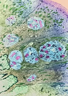
Background imageSpore Collection: Enterocytozoon sp. parasites, TEM

Enterocytozoon sp. parasites, TEM

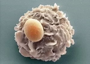
Background imageSpore Collection: Phagocytosis of a yeast spore, SEM

Phagocytosis of a yeast spore, SEM
Phagocytosis of a yeast spore. Coloured scanning electron micrograph (SEM) of a neutrophil (brown) phagocytosing (engulfing and destroying) a spore from the yeast Candida albicans (orange)

Background imageSpore Collection: Phagocytosis of a fungus spore, SEM

Phagocytosis of a fungus spore, SEM
Phagocytosis of a fungus spore. Coloured scanning electron micrograph (SEM) of a spore from the fungus Aspergillus fumigatus (red) being phagocytosed (engulfed and destroyed)

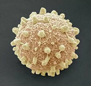
Background imageSpore Collection: Puffball fungus spore, SEM

Puffball fungus spore, SEM
Puffball fungus spore, coloured scanning electron micrograph (SEM). This is the reproductive cell of the fungus. Magnification: x3000 when printed at 10 centimetres wide

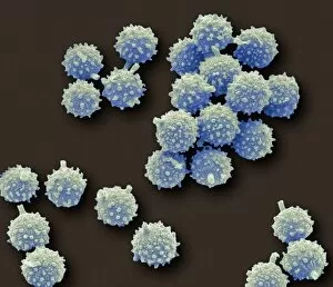
Background imageSpore Collection: Puffball fungus spores, SEM

Puffball fungus spores, SEM
Puffball fungus spores, coloured scanning electron micrograph (SEM). These are the reproductive cells of the fungus. Magnification: x3000 when printed at 10 centimetres wide

Background imageSpore Collection: Pear rust fungus, light micrograph

Pear rust fungus, light micrograph
Pear rust fungus. Light micrograph of a section through tissue from a juniper tree infected with pear rust fungus (Gymnosporangium sabinae, lower right)

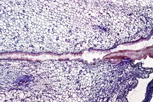
Background imageSpore Collection: Grey mould fungus, light micrograph

Grey mould fungus, light micrograph
Grey mould fungus. Light micrograph of a section through onion tissue infected with grey mould fungus (Botrytis allii). Botrytis is a parasitic saprophyte mould that grows on wounded plants

Background imageSpore Collection: Corn smut infection, light micrograph

Corn smut infection, light micrograph
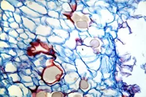
Corn smut infection. Light micrograph of a section through tissue from a corn (Zea mays) plant infected with corn smut fungus (Ustilago maydis)

Background imageSpore Collection: Bramble rust fungus spores

Bramble rust fungus spores. Light micrograph of teliospores from the bramble rust fungus (Phragmidium violaceum). Magnification: x37 when printed 10 centimetres wide

Background imageSpore Collection: Rose mildew fungus, light micrograph

Rose mildew fungus, light micrograph
Rose mildew fungus. Light micrograph of a section through tissue from a rose plant infected with rose mildew fungus (Erysiphe pannosa)

Background imageSpore Collection: Polypody fern sori (Polypodium vulgare)

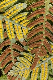
Polypody fern sori (Polypodium vulgare)
Sori on the underside of fronds of the common polypody fern, Polypodium vulgare. The life cycle of a fern consists of alternating generations of which the green fern " plant" is the asexual

Background imageSpore Collection: Black scab infection, light micrograph

Black scab infection, light micrograph
Black scab infection. Light micrograph of a section through a potato infected with black scab (Synchytrium endobioticum) fungus

Background imageSpore Collection: Powdery scab infection, light micrograph

Powdery scab infection, light micrograph
Powdery scab infection. Light micrograph of a section through a potato infected with powdery scab (Spongospora subterranea), showing pustules and spores. S

Background imageSpore Collection: Plum pocket infection, light micrograph

Plum pocket infection, light micrograph
Plum pocket infection. Light micrograph of a section through a plum gall caused by the plum pocket fungus (Taphrina pruni)

Background imageSpore Collection: Rust fungus in a leaf, light micrograph

Rust fungus in a leaf, light micrograph
Rust fungus in a leaf. Light micrograph of a section through a wheat leaf infected with the parasitic Puccinia graminis rust

Background imageSpore Collection: Penicillium mould, light micrograph

Penicillium mould, light micrograph
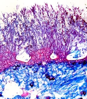
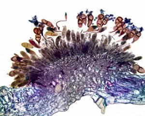
Penicillium mould. Light micrograph of a section through an orange rind infected with Penicillium sp. blue mould fungus, showing fungal hyphae and conidiophores (pink and purple) with spores (red)
Background imageSpore Collection: Brown mould fungus, light micrograph

Brown mould fungus, light micrograph
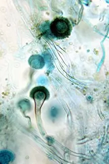
Brown mould fungus. Light micrograph of a section through decaying plant tissue infected with brown mould fungus (Aspergillus glaucum)

Background imageSpore Collection: Downy mildew infection, light micrograph

Downy mildew infection, light micrograph
Downy mildew infection. Light micrograph of a section through a grapevine (Vitis sp.) leaf infected with downy mildew (Plasmopara viticola)

Background imageSpore Collection: Hollyhock rust fungus in a leaf

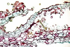
Hollyhock rust fungus in a leaf. Light micrograph of a section through a hollyhock (Alcea sp.) leaf infected with the parasitic Puccinia malvacearum rust, showing the teliospores (spores)

Background imageSpore Collection: Bacterial endospore formation, diagram

Bacterial endospore formation, diagram. The initial stage is at upper left, following the arrow to a ninth stage at lower right

Background imageSpore Collection: Fern spore cases, SEM

Fern spore cases, SEM
Fern spore cases. Coloured scanning electron micrograph (SEM) of a sorus on the underside of a fern leaf. A sorus is a group of sporangia (round lumps)

Background imageSpore Collection: Stilton cheese, SEM

Stilton cheese, SEM
Stilton. Coloured scanning electron micrograph (SEM) of a blue vein in a piece of Stilton cheese. The colour and flavour of the cheese is produced by the fungus Penicillium roquefortii

Background imageSpore Collection: Fern leaf, SEM

Fern leaf, SEM
Fern leaf. Coloured scanning electron micrograph (SEM) of the underside of a fern leaf, showing sori. A sorus is a group of sporangia (brown)

Background imageSpore Collection: Nematode-killing fungus, SEM

Nematode-killing fungus, SEM
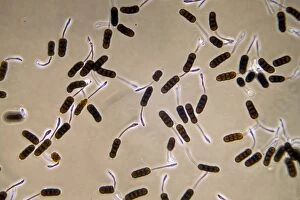
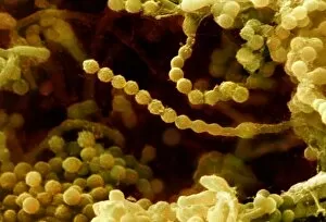
Nematode-killing fungus. Coloured scanning electron micrograph (SEM) of spore chains from the fungus Paecilomyces variotii. This fungus is found in soil and decaying plant matter

Background imageSpore Collection: Fern frond, light micrograph

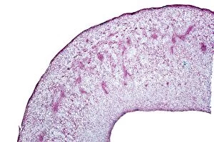
Fern frond, light micrograph. Transverse section through the frond of a fern (Cyrtomium falcatum), showing the sporangia. At left is a central vascular bundle (meristele)

Background imageSpore Collection: Horsetail cone, light micrograph

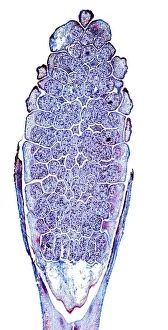
Horsetail cone, light micrograph. Longitudinal section through a young cone (strobilus) of the rough horsetail (Equisetum hyemale)

Background imageSpore Collection: Liverwort spore capsule, light micrograph

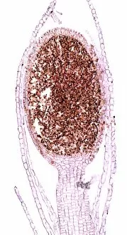
Liverwort spore capsule, light micrograph. Longitudinal section through the thallus and sporangium of a liverwort (Jungammania sp.) The sporangium (spore capsule)

Background imageSpore Collection: Liverwort spore elaters, light micrograph

Liverwort spore elaters, light micrograph. Transverse section through the sporangium of a liverwort (Pellia epiphylla). This is the basal portion of the sporangium

Background imageSpore Collection: Moss spore capsule, light micrograph

Moss spore capsule, light micrograph
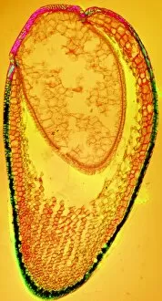
Moss spore capsule, polarised light micrograph. Longitudinal section through a spore capsule from a fire moss (Funaria hygrometrica)


For sale as Licensed Images

Choose your image, Select your licence and Download the media

"Exploring the Fascinating World of Spores: From Aspergillus to Fungal Kingdom" Delving into the microscopic realm, we uncover the enchanting world of spores. Take a closer look at these tiny wonders that play a significant role in various organisms' life cycles. Intriguingly, Aspergillus reveals its intricate structure under scrutiny. Its delicate filaments intertwine, forming an elaborate network that aids in reproduction and dispersal. Moving on to moss capsules like Homalothecium sericeum, scanning electron microscopy (SEM) unveils their mesmerizing architecture. These capsules serve as nature's ingenious vessels for carrying spores, ensuring their propagation across vast distances. Witnessing phagocytosis of fungal spores through SEM is truly captivating. This process showcases how cells engulf these minuscule entities, highlighting the complex interactions between fungi and other organisms within ecosystems. Cyanobacteria also make an appearance under SEM's watchful eye. Their vibrant colors come alive as we observe these photosynthetic microorganisms responsible for oxygen production and nitrogen fixation. The horsetail family presents its own unique spore structures captured by SEM. The common horsetail displays its distinctive spore arrangement while field horsetail exhibits its elegant beauty up close - both showcasing nature's remarkable diversity. Bread mould takes center stage with SEM revealing its intricate filamentous hyphae adorned with countless reproductive spores. These resilient structures ensure bread mould's survival even in unfavorable conditions. Further exploring fungal realms through SEM unravels more astonishing details about their diverse forms and functions. Each species boasts distinct characteristics that aid them in colonizing new habitats or spreading far and wide across landscapes. Clostridium tetani offers us a glimpse into history with an ancient lithograph depicting colonies brimming with dormant yet potent spores dating back to 1906 – reminding us of science's enduring quest to understand the microbial world.