Spore Collection (#7)

"Exploring the Fascinating World of Spores: From Aspergillus to Fungal Kingdom" Delving into the microscopic realm, we uncover the enchanting world of spores

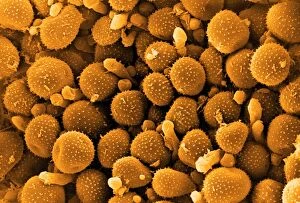
Background imageSpore Collection: Bean rust spores, SEM

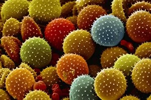
Bean rust spores, SEM
Bean rust (Uromyces sp.) spores, coloured scanning electron micrograph (SEM). Bean rust is a fungal disease that infects bean plants

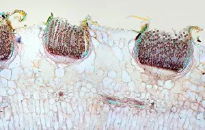
Background imageSpore Collection: Rust fungus infection, light micrograph

Rust fungus infection, light micrograph. Transverse section through a barberry (Berberis sp.) leaf infected by a parasitic rust (Puccinia sp.)

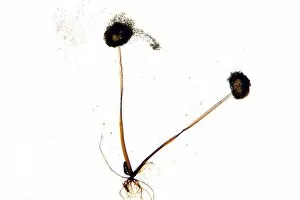
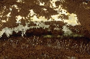
Background imageSpore Collection: Mould fruiting bodies, light micrograph

Mould fruiting bodies, light micrograph. Black mould (Rhizopus nigricans) fruiting bodies (top and upper right) liberating spores

Background imageSpore Collection: Aspergillus fungus spores, SEM

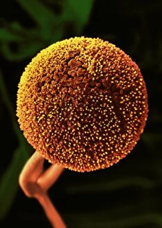
Aspergillus fungus spores, SEM
Aspergillus fungus spores, coloured scanning electron micrograph (SEM). These chains of spores (orange) are the reproductive cells of the fruiting body, or conidiophore

Background imageSpore Collection: Penicillium fungus, SEM

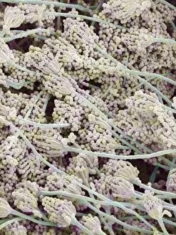
Penicillium fungus, SEM
Penicillium fungus. Coloured scanning electron micrograph (SEM) of a Penicillium sp. fungus. Specialised threads (hyphae, pink), called conidiophores, are seen

Background imageSpore Collection: Coloured SEM of anthrax bacteria spores

Coloured SEM of anthrax bacteria spores

Background imageSpore Collection: Clostridium tetani bacterial spore

Clostridium tetani bacterial spore
False-colour transmission electron micrograph (TEM) of a Clostridium tetani bacterial spore. The bacterium causes tetanus (lockjaw)

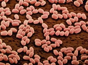
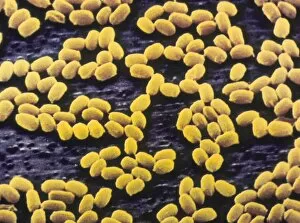
Background imageSpore Collection: Anthrax bacteria spores

Anthrax bacteria spores
False-colour scanning electron micrograph of spores of the large, rod-shaped bacteria Bacillus anthracis, the causative agent of anthrax in farm animals

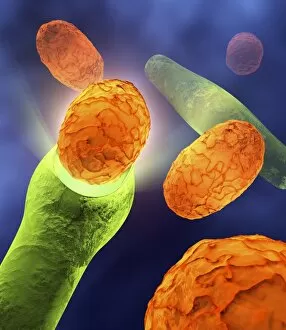
Background imageSpore Collection: Anthrax bacteria

Anthrax bacteria. Computer artwork of Bacillus anthracis bacteria (green) releasing spores (orange). These are Gram-positive, rod-shaped, spore-forming bacteria

Background imageSpore Collection: Liverwort spore case, light micrograph

Liverwort spore case, light micrograph
Liverwort spore case. Light micrograph of a vertical section through a spore case (sporophyte) from a liverwort (Pellia epiphylla)

Background imageSpore Collection: Bracken spores, SEM

Bracken spores, SEM
Bracken spores. Coloured scanning electron micrograph (SEM) of the leaves of a bracken plant (Pteridium aquilinum). The undersides of the leaves are covered in spore-containing sacs known as

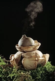
Background imageSpore Collection: Earth star mushroom

Earth star mushroom (Geastrum triplex) growing in woodland soil. It has released (puffed) a cloud of reproductive spores (upper centre)

Background imageSpore Collection: Horsetail spores, SEM

Horsetail spores, SEM
Horsetail spores. Coloured scanning electron micrograph (SEM) of spores of a horsetail (Equisetum sp.). The spores (orange) bear elaters (green), which expand and contract with changes in humidity

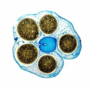
Background imageSpore Collection: Liverwort spore cases, light micrograph

Liverwort spore cases, light micrograph
Liverwort spore cases. Light micrograph of a cross-section through five spore cases (sporophytes) from the liverwort Conocephalum conicum (Marchantiales)

Background imageSpore Collection: Fern spores, SEM

Fern spores, SEM
Fern spores. Coloured scanning electron micrograph (SEM) of spores (red) of a fern partly enclosed in a sporangium. The undersides of the leaves of ferns are covered in these spore-containing sacs

Background imageSpore Collection: Bracken leaves, SEM

Bracken leaves, SEM
Bracken leaves. Coloured scanning electron micrograph (SEM) of the leaves of a bracken plant (Pteridium aquilinum). The undersides of the leaves are covered in spore-containing sacs known as

Background imageSpore Collection: Sporangium of bread mould, Rhizopus

Sporangium of bread mould, Rhizopus
False colour scanning electron micrograph of a sporangium, or fruiting body, of the common bread mould, Rhizopus stolonifer. Spores circulate freely in the air

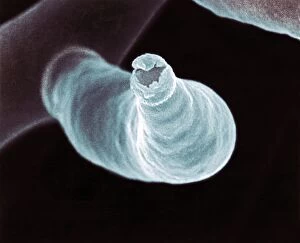
Background imageSpore Collection: Fungal reproduction, SEM

Fungal reproduction, SEM
Fungal reproduction. Coloured scanning electron micrograph (SEM) of the tip of a branch of a conidiophore (one type of fungal reproductive structure) with a conidia (spore) emerging from the tip

Background imageSpore Collection: Spore of field horsetail

Spore of field horsetail
False colour scanning electron micrograph of spores of the field horsetail, Equisetum arvense. The horsetail is the only living type of a primitive family of plants, the Sphenopsids

Background imageSpore Collection: Bread mould, Rhizopus

Bread mould, Rhizopus
Macrophotograph of a thirteen day old growth of a bread mould (fungus) Rhizopus sp. The slices of bread are overgrown with a network of vegetative hyphae (transparent & threadlike)

Background imageSpore Collection: Northern moonwort (Botrychium boreale)

Northern moonwort (Botrychium boreale) with spores (small round specks) on its fertile fronds. Photographed in Norway

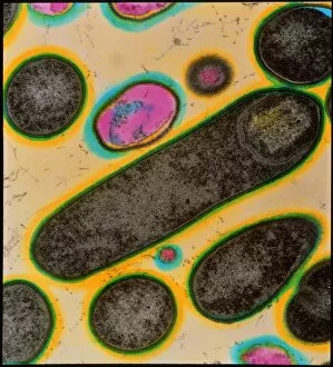
Background imageSpore Collection: Clostridium bacteria

Clostridium bacteria
Coloured transmission electron micrograph (TEM) of Clostridium botulinum bacteria, the cause of botulism food poisoning. The bacterium seen in longitudinal section at centre is in the process of

Background imageSpore Collection: Clostridium botulinum bacteria with spores

Clostridium botulinum bacteria with spores
Coloured transmission electron micrograph (TEM) of the bacteria Clostridium botulinum, cause of botulism food poisoning in humans, in the process of sporulation (spore formation)

Background imageSpore Collection: Clostridium difficile bacteria, TEM

Clostridium difficile bacteria, TEM
Clostridium difficile drug-resistant bacteria, coloured transmission electron micrograph (TEM). This bacterium, also known as C. diff, is a normal member of the intestinal flora in humans

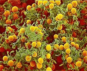
Background imageSpore Collection: Athletes foot fungus, SEM

Athletes foot fungus, SEM
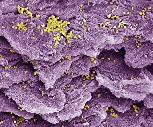
Athletes foot. Coloured scanning electron micrograph (SEM) of spores (yellow) of the fungus that causes athletes foot (tinea pedis) on skin (purple) from a human foot

Background imageSpore Collection: Stiff clubmoss (Lycopodium annotinum)

Stiff clubmoss (Lycopodium annotinum) in a pine forest. Also called interrupted clubmoss, this primitive plant is commonly found in moist forest, thickets

Background imageSpore Collection: Wood horsetail (Equisetum sylvaticum)

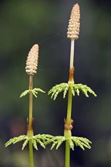
Wood horsetail (Equisetum sylvaticum). This plant grows in moist, cool woods and has many delicate branches that circle the shoots. Photographed in Scotland

Background imageSpore Collection: Male giant horsetail (Equisetum arvense)

Male giant horsetail (Equisetum arvense)
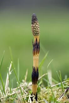
Mail giant horsetail (Equisetum telemataia) with a fertile cone at the tip of its stem. Photographed in spring

Background imageSpore Collection: Common horsetail (Equisetum arvense)

Common horsetail (Equisetum arvense)
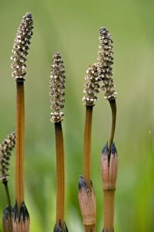
Common horsetail plant (Equisetum arvense) with fertile cones. Photographed in spring

Background imageSpore Collection: Fern spore cases, light micrograph

Fern spore cases, light micrograph
Fern spore cases. Polarised light micrograph of a frond of the maidenhair fern (Adiantum capillus- veneris), showing the sporangia (black, centre left) which contain the ferns spores

Background imageSpore Collection: Flowering fern spores, SEM

Flowering fern spores, SEM
Flowering fern spores. Coloured scanning electron micrograph (SEM) of three spores of the flowering fern (Osmunda regalis). The flowering, or royal, fern, is found across much of the world

Background imageSpore Collection: Rust fungus spores, SEM

Rust fungus spores, SEM
Rust fungus spores. Coloured scanning electron micrograph (SEM) of spores from a rust fungus that infects rose plants. Rust fungi (order Uredinales) are parasites that attack plants

Background imageSpore Collection: Mushroom, light micrograph

Mushroom, light micrograph
Mushroom. Light micrograph (LM) of a section through a mushroom, Agaricus sp. (formerly Psalliota sp.), showing its stalk (orange) and gills (long white projections)

Background imageSpore Collection: Mushroom gills, light micrograph

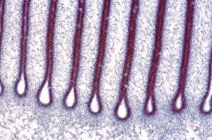
Mushroom gills, light micrograph
Mushroom gills. Light micrograph (LM) of a section through the gills of a mushroom, Agaricus sp. (formerly Psalliota sp.)

Background imageSpore Collection: Bread mould

Bread mould (Penicillium sp.). Coloured scanning electron micrograph (SEM) of mouldy bread. The fungus penetrates the bread, digesting and absorbing its nutrients

Background imageSpore Collection: Bread mould, SEM

Bread mould, SEM
Bread mould. Coloured scanning electron micrograph (SEM) of a sporangium of a bread mould (Rhizopus stolonifer). Spores develop in these sac-like bodies, or sporangia (globular structure)
Background imageSpore Collection: USA, Oregon, Treefrog

USA, Oregon, Treefrog on Morel

Background imageSpore Collection: Honey Fungus - caps with spores, on autumn leaf litter, Hessen, Germany

Honey Fungus - caps with spores, on autumn leaf litter, Hessen, Germany
USH-3757 Honey Fungus - caps with spores, on autumn leaf litter Hessen, Germany Armillariella mellea Duncan Usher Please note that prints are for personal display purposes only

Background imageSpore Collection: Spores on the uderside of a fern

Spores on the uderside of a fern
Ashley Cooper / SpecialistStock

Background imageSpore Collection: Bread mould (Penicillium sp. ), SEM

Bread mould (Penicillium sp. ), SEM
Bread mould. Coloured scanning electronmicrograph (SEM) of Penicillium sp. mouldgrowing on bread. The yellow fibrous structuresare hyphae, which make up the main body of thefungus


For sale as Licensed Images

Choose your image, Select your licence and Download the media

"Exploring the Fascinating World of Spores: From Aspergillus to Fungal Kingdom" Delving into the microscopic realm, we uncover the enchanting world of spores. Take a closer look at these tiny wonders that play a significant role in various organisms' life cycles. Intriguingly, Aspergillus reveals its intricate structure under scrutiny. Its delicate filaments intertwine, forming an elaborate network that aids in reproduction and dispersal. Moving on to moss capsules like Homalothecium sericeum, scanning electron microscopy (SEM) unveils their mesmerizing architecture. These capsules serve as nature's ingenious vessels for carrying spores, ensuring their propagation across vast distances. Witnessing phagocytosis of fungal spores through SEM is truly captivating. This process showcases how cells engulf these minuscule entities, highlighting the complex interactions between fungi and other organisms within ecosystems. Cyanobacteria also make an appearance under SEM's watchful eye. Their vibrant colors come alive as we observe these photosynthetic microorganisms responsible for oxygen production and nitrogen fixation. The horsetail family presents its own unique spore structures captured by SEM. The common horsetail displays its distinctive spore arrangement while field horsetail exhibits its elegant beauty up close - both showcasing nature's remarkable diversity. Bread mould takes center stage with SEM revealing its intricate filamentous hyphae adorned with countless reproductive spores. These resilient structures ensure bread mould's survival even in unfavorable conditions. Further exploring fungal realms through SEM unravels more astonishing details about their diverse forms and functions. Each species boasts distinct characteristics that aid them in colonizing new habitats or spreading far and wide across landscapes. Clostridium tetani offers us a glimpse into history with an ancient lithograph depicting colonies brimming with dormant yet potent spores dating back to 1906 – reminding us of science's enduring quest to understand the microbial world.