Stigma Collection (#7)

"Breaking the Chains: Unraveling the Stigma Surrounding Nature's Wonders" In Saint Joseph of Cupertino's Ecstasy

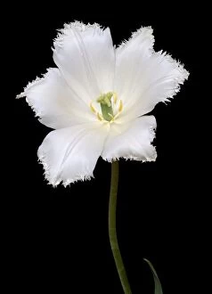
Background imageStigma Collection: Tulip (Tulipa gesneriana), hybrid form C010 / 7011

Tulip (Tulipa gesneriana), hybrid form C010 / 7011
Tulip (Tulipa gesneriana) flowering in Spring, showing the stigma and stamens

Background imageStigma Collection: Christs stigmata, 17th century

Christs stigmata, 17th century
Christs stigmata. Artwork of Christs rope-bound hands, showing the site of the stigmata. In this context, stigmata refers to the Holy Wounds that Jesus Christ received during his crucifixion

Background imageStigma Collection: 1862 Darwins slipper orchid pollination

1862 Darwins slipper orchid pollination
Composite of Darwins illustration of Cypripedium (slipper orchid, Paphiopedilum) below and a matched photograph of an early variety of Sandford orchid cultivar above

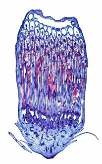
Background imageStigma Collection: Broad bean flower, light micrograph

Broad bean flower, light micrograph
Broad bean flower. Polarised light micrograph of a section through a flower from a broad bean (Vicia faba) plant, showing the stamens and stigma

Background imageStigma Collection: Dandelion flower, light micrograph

Dandelion flower, light micrograph
Dandelion flower. Light micrograph of a section through a flower from a dandelion (Taraxacum officinalis) plant. The flower stem (bottom) is made up of parenchyma cells (light blue)

Background imageStigma Collection: Shepherds purse fruit, light micrograph

Shepherds purse fruit, light micrograph
Shepherds purse fruit. Light micrograph of a section through a seed pod from a shepherds purse (Capsella bursa-pastoris) plant. The remains of the stigma can be seen (top centre)

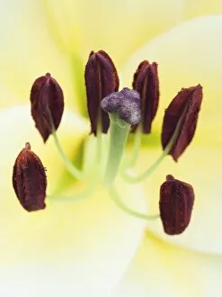
Background imageStigma Collection: Lily reproductive parts

Lily reproductive parts. Close-up of the stamens and carpel of a hybrid garden lily. Lilies display three-fold symmetry in their floral parts

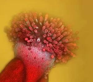
Background imageStigma Collection: Thale cress flower, micrograph

Thale cress flower, micrograph
Thale cress flower. Fluorescence micrograph of a flower from a thale cress (Arabidopsis thaliana) plant. The female reproductive structures - carpel (red) and stigma (yellow) - can be seen at centre

Background imageStigma Collection: Thale cress stigma, micrograph

Thale cress stigma, micrograph
Thale cress stigma. Fluorescence micrograph of a stigma (female reproductive structure) from a thale cress (Arabidopsis thaliana) flower

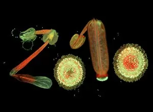
Background imageStigma Collection: Stamens and carpels, micrograph

Stamens and carpels, micrograph
Stamens and carpels. Fluorescence micrograph of stamens (male reproductive structures, far left) and carpels (female reproductive structures)

Background imageStigma Collection: Thale cress stigma, micrograph

Thale cress stigma, micrograph
Thale cress stigma. Fluorescence micrograph of a stigma (female reproductive structure) from a thale cress (Arabidopsis thaliana) flower

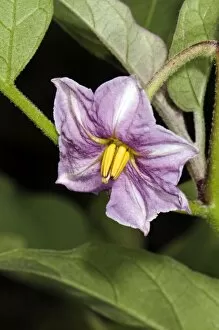
Background imageStigma Collection: Aubergine (Solanum melongena) flower

Aubergine (Solanum melongena) flower. Close-up of a flower on an aubergine plant. Photographed in Orvieto, Umbria, Italy

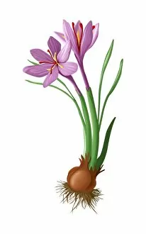
Background imageStigma Collection: Saffron flowers and bulb

Saffron flowers and bulb. The flowers (pink) have grown from the bulb (bottom). Also seen are the roots and the leaves (green). The part of the plant most commonly used is the stigmas (red)

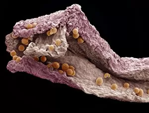
Background imageStigma Collection: Saffron, SEM

Saffron, SEM
Saffron, coloured scanning electron micrograph (SEM). Saffron is a spice derived from the dried stigmas (orange) of the saffron crocus flower (Crocus sativus)

Background imageStigma Collection: Euglena protozoan, light micrograph

Euglena protozoan, light micrograph
Euglena protozoan. Differential interference contrast micrograph of the flagellate protozoan Euglena spirogyra. This freshwater single-celled organism can either obtain energy from sunlight

Background imageStigma Collection: Euglena protozoa, light micrograph

Euglena protozoa, light micrograph
Euglena protozoa. Differential interference contrast micrograph of the flagellate protozoa Euglena acus. These freshwater single-celled organisms can either obtain energy from sunlight

Background imageStigma Collection: Small scabious (Scabiosa columbaria)

Small scabious (Scabiosa columbaria) in flower (centre), with a fruit (left). This plant, found on chalk downlands, is also known as doves foot scabious. Photographed in Hampshire, England

Background imageStigma Collection: Glacier lily (Erythronium grandiflorum)

Glacier lily (Erythronium grandiflorum) flowering in the snow. Photographed on Mount Rainier, Cascade Mountains, Washington, USA

Background imageStigma Collection: Hypericum stigma with pollen grains, SEM

Hypericum stigma with pollen grains, SEM
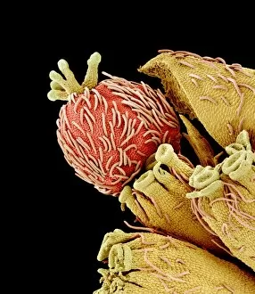
Hypericum stigma (brown) with pollen grains (pink and yellow), coloured scanning electron micrograph (SEM). Pollen grains are reproductive structures produced by the male parts of flowering plants

Background imageStigma Collection: Starry saxifrage (Saxifraga stellaris)

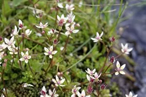
Starry saxifrage (Saxifraga stellaris) in flower. Photographed in the Cairngorms National Park, Scottish Highlands, in June

Background imageStigma Collection: Tulip (Tulipa pulchella)

Tulip (Tulipa pulchella) flower. Close-up of the centre of a tulip flower showing the reproductive parts. Six stamens can be seen in a ring around the stigma (pale yellow)

Background imageStigma Collection: Cypriot tulip flower (Tulipa cypria)

Cypriot tulip flower (Tulipa cypria). This plant is endemic to Cyprus. Photographed in the Akamas peninsula in Cyprus

Background imageStigma Collection: Leopard lily (Lilium pardalinum)

Leopard lily (Lilium pardalinum) flowers. Photographed on Mount Eddy, Klamath Mountains, northern California, USA

Background imageStigma Collection: Crocus tommasinianus

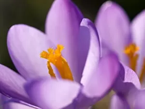
Crocus tommasinianus in flower in February, showing the stigma with its brush-like end and the pollen covered stamens around it

Background imageStigma Collection: Ladys mantle reproductive parts, SEM

Ladys mantle reproductive parts, SEM
Ladys mantle reproductive parts, coloured scanning electron micrograph (SEM). Ladys mantle, a plant in the genus Alchemilla

Background imageStigma Collection: Drifts of Crocuses naturalised in grass

Drifts of Crocuses naturalised in grass
Drifts of Crocus tommasinianus naturalised in grass, a garden lawn in Dorset, UK. These are some of the first spring flowers in many gardens (late February into early March in the UK)

Background imageStigma Collection: Morning glory pollination, SEM

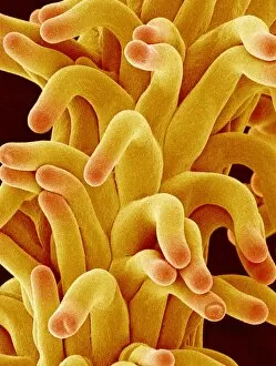
Morning glory pollination, SEM
Morning glory pollination. Coloured scanning electron micrograph (SEM) of a morning glory (Ipomoea sp.) flower pistil (fringed lobes) with attached pollen grains (round)

Background imageStigma Collection: Caper flower and buds

Caper flower and buds
Caper plant (Capparis spinosa) in flower, with an open flower (centre) and several unopened flower buds (green). This is a spiny shrub, native to Mediterranean and Caucasian areas

Background imageStigma Collection: Euphorbia flower reproductive parts, SEM

Euphorbia flower reproductive parts, SEM
Euphorbia flower reproductive parts. Coloured scanning electron micrograph (SEM) of the reproductive parts of a Euphorbia supina flower

Background imageStigma Collection: Sunflower pollination, SEM

Sunflower pollination, SEM
Sunflower pollination. Coloured scanning electron micrograph (SEM) of pollen on a stigma of a sunflower plant (Helianthus sp.)

Background imageStigma Collection: Sweet pea flower pollination, SEM

Sweet pea flower pollination, SEM
Sweet pea pollination. Coloured scanning electron micrograph (SEM) of pollen grains (yellow) on the stigma (green) of a sweet pea flower (Lathyrus odoratus)

Background imageStigma Collection: SEM of pollen germinating stigma of turnip flower

SEM of pollen germinating stigma of turnip flower
False-colour scanning electron micrograph (SEM) of pollen germinating on the stigma of the turnip flower, Brassica campestris

Background imageStigma Collection: Passion flower

Passion flower (Passiflora Venus ). The passion flower is a tropical climbing plant that is native to South America. The flower consists of white petals

Background imageStigma Collection: Thunberg spirea, SEM

Thunberg spirea, SEM
Thunberg spirea (Spiraea thunbergii). Coloured scanning electron micrograph (SEM) of a flower of Thunberg spirea, a member of the rose family, found in China and Japan

Background imageStigma Collection: Morning glory flower pollination, SEM

Morning glory flower pollination, SEM
Morning glory flower pollination. Coloured scanning electron micrograph (SEM) of pollen grains (orange) on the stigma of a morning glory flower (Ipomoea learii)

Background imageStigma Collection: Enkianthus flower, SEM

Enkianthus flower, SEM
Enkianthus flower. Coloured scanning electron micrograph (SEM) of the reproductive structures inside an Enkianthus perulatus flower

Background imageStigma Collection: Easter cactus stigma, SEM

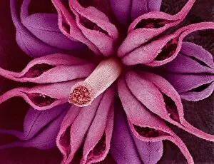
Easter cactus stigma, SEM
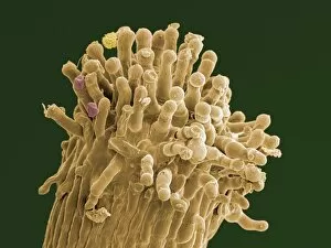
Easter cactus stigma. Coloured scanning electron micrograph (SEM) of part of the stigma (pink) of an Easter cactus flower (Rhipsalidopsis gaertneri)

Background imageStigma Collection: Pollination, light micrograph

Pollination, light micrograph
Pollination. Light micrograph of a section through an evening primrose (Oenthera biennis) stigma. Pollen grains (circular) are adhering to the surface of the stigma

Background imageStigma Collection: Wild strawberry flower pistil, SEM

Wild strawberry flower pistil, SEM
Wild strawberry pistil. Coloured scanning electron micrograph (SEM) of the pistil of a flower of the Asian wild strawberry (Potentilla sp.). The pistil is the female reproductive part of a flower

Background imageStigma Collection: Germination of turnip pollen

Germination of turnip pollen

Background imageStigma Collection: Pollen germinating on stigma of goosegrass

Pollen germinating on stigma of goosegrass

Background imageStigma Collection: Chrysanthemum pistil, SEM

Chrysanthemum pistil, SEM
Chrysanthemum pistil. Coloured scanning electron micrograph (SEM) of a pistil from a Chrysanthemum sp. flower. The pistil is the female reproductive part of a plant

Background imageStigma Collection: Zinnia disc floret, SEM

Zinnia disc floret, SEM
Zinnia disc floret. Coloured scanning electron micrograph (SEM) of a Zinnia sp. flower head disc floret. This floret (true flower) is found in the central (disc) region of the flower head

Background imageStigma Collection: Passion flowers

Passion flowers (Passiflora Venus ). The passion flower is a tropical climbing plant that is native to South America. The flower consists of white petals

Background imageStigma Collection: Chickweed flower pistil, SEM

Chickweed flower pistil, SEM
Chickweed flower pistil. Coloured scanning electron micrograph (SEM) of a chickweed flower (Stellaria media) pistil. Pistils hold a plants female reproductive structures

Background imageStigma Collection: Pollen on stigma of Cocksfoot grass

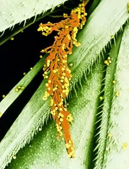
Pollen on stigma of Cocksfoot grass
False-colour scanning electron micrograph (SEM) of pollen grains clustered to the stigma of the cocksfoot grass, Dactylis glomerata

Background imageStigma Collection: Morning glory pollen

Morning glory pollen grains. Coloured scanning electron micrograph (SEM) of pollen grains (orange balls) on the pistil (female reproductive parts) of a morning glory flower (Ipomoea sp.)

Background imageStigma Collection: Chickweed flower pistil and pollen, SEM

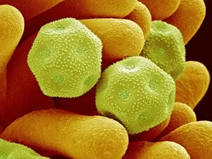
Chickweed flower pistil and pollen, SEM
Chickweed flower pistil and pollen. Coloured scanning electron micrograph (SEM) of a chickweed flower (Stellaria media) pistil (yellow) and pollen grains (green)


For sale as Licensed Images

Choose your image, Select your licence and Download the media

"Breaking the Chains: Unraveling the Stigma Surrounding Nature's Wonders" In Saint Joseph of Cupertino's Ecstasy, we witness a divine connection that transcends human understanding. Just like this masterpiece, stigma in nature holds profound significance. The Methodus plantarum sexalis in sistemate naturae descripta reveals the intricate classification of plants, shedding light on how stigma plays a vital role in their reproductive processes, and is through these delicate structures that life perpetuates itself. Zooming into an Easter cactus stigma under SEM, we discover its mesmerizing beauty up close. The Studio shot captures the essence of a pink tulip flower with such precision that we can almost feel its velvety touch. Morning glory pollen captured by SEM showcases the interplay between stigma and pollen grains—a dance essential for successful pollination. Similarly, Gorse stigma adorned with pollen grains exemplifies nature's harmonious collaboration to ensure survival. A False-colour SEM image of chickweed flower reminds us that even seemingly insignificant flora possess unique stigmas worthy of admiration. Each microscopic detail tells a story waiting to be unraveled. Pollination takes center stage as different species intertwine their destinies through this sacred union. Amaryllis stands tall as Hippeastrum sp. , showcasing vibrant stigmas inviting pollinators to partake in their splendorous feast. Traveling to Greece, we encounter Saffron Crocus—its precious saffron harvested from meticulously hand-picked stigmas—an ancient tradition steeped in culture and history. Lastly, Thale cress flower micrograph serves as a testament to scientific exploration; it invites us into its miniature world where every aspect contributes to our understanding of life's intricacies. Through these captivating glimpses into nature's wonders, let us challenge societal norms and break free from the chains of prejudice surrounding "stigma. " May we recognize its inherent beauty and importance, both in art and the natural world.