Structures Collection (#58)

"Captivating Structures from Around the World: A Visual Journey" Witness the mesmerizing sunrise behind the majestic Angkor Wat

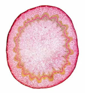
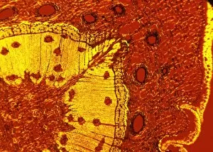
Background imageStructures Collection: Spikenard stem, light micrograph

Spikenard stem, light micrograph
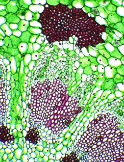
Spikenard stem. Light micrograph of a cross-section through the stem of a Spikenard (Aralia sp.) tree. The dark red cells just beneath the surface bark are collenchyma cells

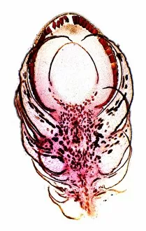
Background imageStructures Collection: Female yew tree flower, light micrograph

Female yew tree flower, light micrograph
Female yew tree flower. Light micrograph of a longitudinal section through a female flower of the yew tree (Taxus baccata)

Background imageStructures Collection: Buttercup flower bud, light micrograph

Buttercup flower bud, light micrograph
Buttercup flower bud. Light micrograph of a cross-section through an immature wallflower bud (Ranunculus sp.), showing the male and female reproductive organs of the flower

Background imageStructures Collection: Flower ovary and ovules, SEM

Flower ovary and ovules, SEM
Flower ovary and ovules. Coloured freeze-fracture scanning electron micrograph (SEM) through the ovary of a dicot flower. The ovary (purple) contains ovules (green)

Background imageStructures Collection: Dog rose stem, light micrograph

Dog rose stem, light micrograph
Dog rose stem. Polarised light micrograph of a cross-section through the stem of a dog rose (Rosa canina) plant. The outer cortex (black and red) is made of cork cambium tissue

Background imageStructures Collection: Alexander flowers, light micrograph

Alexander flowers, light micrograph
Alexander flowers. Light micrograph of a section through a cluster of alexander flowers (Smyrnium olusatrum). Seen here are cross-sections of the central umbels

Background imageStructures Collection: Elm stem, light micrograph

Elm stem, light micrograph
Elm stem (Ulmus procera) section, polarised light micrograph. The black horizontal lines are multiseriate rays, which are composed of several rows of cells

Background imageStructures Collection: Cucumber flower bud, light micrograph

Cucumber flower bud, light micrograph
Cucumber flower bud. Light micrograph of a longitudinal section through the flower bud of a cucumber (Cucurbita sp.). The female pistil (red)

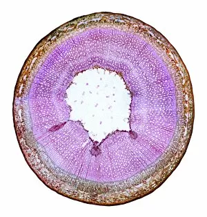
Background imageStructures Collection: Willow stem, light micrograph

Willow stem, light micrograph
Willow stem. Light micrograph of a cross-section through the stem of a willow (Salix sp.) tree. At centre is the pith (white), surrounded by woody xylem tissue (pink)

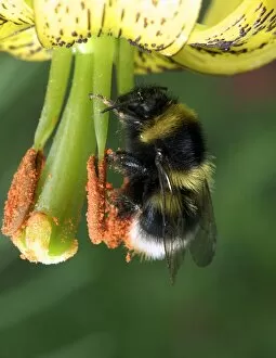
Background imageStructures Collection: Bumblebee collecting pollen

Bumblebee collecting pollen. Close-up of a bumblebee (Bombus sp.) collecting pollen from the stamens of a flower

Background imageStructures Collection: Hydrozoan colony

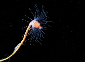
Hydrozoan colony. Close-up of the tentacled feeding structures of a hydrozoan (Tubularia larynx) colony. Hydrozoa are a type of cnidarian and are related to corals and sea anemones

Background imageStructures Collection: Nudibranch eating hydrozoa

Nudibranch eating hydrozoa
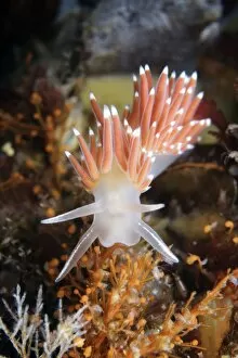
Nudibranch (Flabellina verrucosa) eating hydrozoa (Eudendrium sp.). Nudibranchs are shell-less marine molluscs that live in seas around the world

Background imageStructures Collection: Hydrozoan colony and amphipods

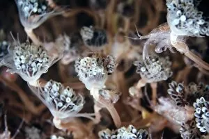
Hydrozoan colony and amphipods. Amphipod (Stenothoe marina) crustaceans sheltering amongst the tentacled feeding structures of a hydrozoan (Tubularia larynx) colony

Background imageStructures Collection: Hydrozoan

Hydrozoan (Tubularia indivisa). Hydrozoa are a type of cnidarian and are related to corals and sea anemones

Background imageStructures Collection: The Eden Project in Cornwall

The Eden Project in Cornwall
The Glasshouses at The Eden Project in Cornwall, UK

Background imageStructures Collection: Rubber and gutta-percha molecular models

Rubber and gutta-percha molecular models
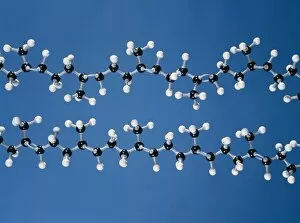
Molecular models of rubber and gutta-percha. These molecules are isomers. They have the same chemical fourmula but a different molecular structure

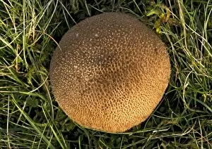
Background imageStructures Collection: Puffball fungus

Puffball fungus (Lycoperdon foetidum)

Background imageStructures Collection: Atlas cedar cones

Atlas cedar cones
Atlas cedar cone (Cedrus atlantica). This is a mature, two-year-old female cone (seed)

Background imageStructures Collection: Calabrian pine (Pinus brutia)

Calabrian pine (Pinus brutia)
Calabrian pine cones (Pinus brutia). Photographed in Greece, in spring

Background imageStructures Collection: Oyster mushrooms

Oyster mushrooms (Pleurotus ostreatus) growing on a tree trunk. These fruiting bodies (reproductive structures) are produced year-round by the fungus in large clusters

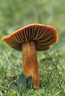
Background imageStructures Collection: Crimson wax cap mushroom

Crimson wax cap mushroom (Hygrocybe punicea). Photographed in the New Forest, UK

Background imageStructures Collection: Lead, SEM

Lead, SEM
Lead, coloured scanning electron micrograph (SEM). Lead is a heavy, soft, ductile metal. It is used in building technology, batteries and bullets

Background imageStructures Collection: Maple stem, light micrograph

Maple stem, light micrograph
Maple stem. Polarised light micrograph of a cross-section through the stem of a field maple (Acer campestre). This shows the thick layer of cork (phellem)

Background imageStructures Collection: Nettle leaf stalk structures, LM

Nettle leaf stalk structures, LM
Nettle leaf stalk structures. Polarised light micrograph (LM) of a nettle leaf (Urtica dioica), which has been boiled up with dilute sodium hydroxide to separate the cells

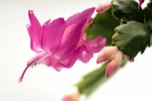
Background imageStructures Collection: Schlumbergera bridgesii

Schlumbergera bridgesii
Christmas cactus flowers (Schlumbergera bridgesii). This cactus is found in the Organ Mountain forests, north of Rio de Janeiro, Brazil. It is cultivated in Europe as a Christmas plant

Background imageStructures Collection: Bogbean stem, light micrograph

Bogbean stem, light micrograph
Bogbean stem. Light micrograph of a cross-section through the stem of a bogbean plant (Menyanthes trifoliata), showing its internal structure

Background imageStructures Collection: Scots pine tree stem, light micrograph

Scots pine tree stem, light micrograph
Scots pine tree stem. Polarised light micrograph of a cross-section through the stem of a Scots pine tree (Pinus sylvestris), showing resin canals

Background imageStructures Collection: Mistletoe vascular bundle, LM

Mistletoe vascular bundle, LM
Mistletoe vascular bundle. Light micrograph (LM) of a section of mistletoe (Viscum album) stem in cross-section, showing a vascular bundle

Background imageStructures Collection: Mistletoe stem, LM

Mistletoe stem, LM
Mistletoe stem. Light micrograph (LM) of a mistletoe (Viscum album) stem in cross-section, showing nine vascular bundles radiating out from the centre of the stem

Background imageStructures Collection: Pansy flower, SEM

Pansy flower, SEM
Pansy flower reproductive parts. Coloured scanning electron micrograph (SEM) of reproductive parts from the flower of a pansy (Viola sp.) plant

Background imageStructures Collection: Mimosa flowers, SEM

Mimosa flowers, SEM
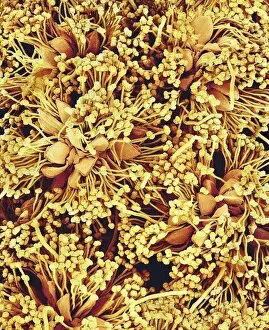
Mimosa flower stamens. Coloured scanning electron micrograph (SEM) of flowers and stamens of Mimosa sp. plants. Many stamens (male reproductive parts) are seen

Background imageStructures Collection: Common pollen beetles feeding

Common pollen beetles feeding
Common pollen beetles (Meligethes aeneus) feeding on pollen from a dog rose flower (Rosa canina)

Background imageStructures Collection: Nigella seed, SEM

Nigella seed, SEM
Nigella seed. Coloured scanning electron micrograph (SEM) of a seed from a nigella flower (family Ranunculaceae). A seed contains an embryo (a fertilised ovule from which a new plant can grow)

Background imageStructures Collection: Roxburgh fig fruit (Ficus auriculata)

Roxburgh fig fruit (Ficus auriculata). Photographed in Asia

Background imageStructures Collection: Creeping zinnia seed, SEM

Creeping zinnia seed, SEM
Creeping zinnia seed. Coloured scanning electron micrograph (SEM) of a seed from a creeping zinnia flower (Sanvitalia sp.)

Background imageStructures Collection: Tobacco, SEM

Tobacco, SEM
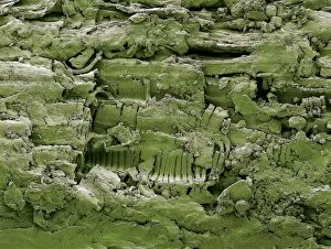
Tobacco. Coloured scanning electron micrograph (SEM) of tobacco from a cigarette. At centre bottom is some spiral xylem tissue

Background imageStructures Collection: Passionflower

Passionflower (Passiflora Adularia )

Background imageStructures Collection: Reproductive flower parts, SEM

Reproductive flower parts, SEM
Reproductive parts of flowers. Coloured scanning electron micrograph (SEM). These are florets, the true flowers that form the flowering structures of plants in the Compositae family

Background imageStructures Collection: Wildflower seed, SEM

Wildflower seed, SEM
Wildflower seed. Coloured scanning electron micrograph (SEM) of a seed from a wildflower. Wildflower is a general term for flowers growing wild in nature

Background imageStructures Collection: Enkianthus flower, SEM

Enkianthus flower, SEM
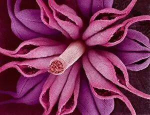
Enkianthus flower. Coloured scanning electron micrograph (SEM) of the reproductive structures inside an Enkianthus perulatus flower

Background imageStructures Collection: Red campion seed, SEM

Red campion seed, SEM
Red campion seed. Coloured scanning electron micrograph (SEM) of a seed from a red campion flower (Silene dioica). A seed contains an embryo (a fertilised ovule from which a new plant can grow)

Background imageStructures Collection: Lily flower reproduction

Lily flower reproduction. Reproductive parts of a lily flower (Lilium sp.). The male stamens consist of a filament topped by a pollen bearing anther (orange)

Background imageStructures Collection: Plant seeds

Plant seeds
Seeds. Coloured scanning electron micrograph (SEM) of seeds. A seed is the product of pollination and is the ripened ovule (egg) of a plant

Background imageStructures Collection: Poppy ovary with developing seeds, LM

Poppy ovary with developing seeds, LM
Poppy ovary with developing seeds. Light micrograph (LM) of a cross-section through the centre of a mature poppy ovary, showing placentas bearing developing seeds (red)

Background imageStructures Collection: Flower pistil, SEM

Flower pistil, SEM
Flower pistil. Coloured scanning electron micrograph (SEM) of the pistil of a tricyrtis (Tricyrtis sp.) flower. The pistil is the female reproductive part of the flower

Background imageStructures Collection: Copihue seed, SEM

Copihue seed, SEM
Copihue seed. Coloured scanning electron micrograph (SEM) of a seed from a copihue, or Chilean glory, flower (Lapageria rosea)

Background imageStructures Collection: European wand loosestrife flowers

European wand loosestrife flowers
European wand loosestrife (Lythrum virgatum) flowers

Background imageStructures Collection: Cherry blossom pollen, SEM

Cherry blossom pollen, SEM
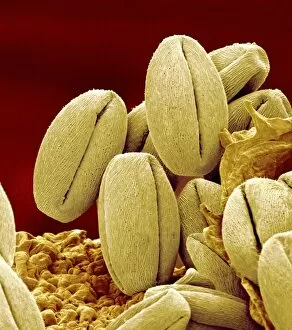
Cherry blossom pollen. Coloured scanning electron micrograph (SEM) of pollen grains from a cherry tree flower (Prunus yedoensis)


For sale as Licensed Images

Choose your image, Select your licence and Download the media

"Captivating Structures from Around the World: A Visual Journey" Witness the mesmerizing sunrise behind the majestic Angkor Wat, a testament to Cambodia's rich history and architectural brilliance in Siem Reap. The grandeur of St Pauls Cathedral in Melbourne is amplified by its magnificent pipe organ, filling the air with enchanting melodies that resonate through time. Discover serenity as you behold a Buddha statue at Borobudur adorned with Dharmachakra Mudra hand position during a breathtaking sunrise in Magelang, Central Java. Gaze upon the stunning panorama of Victoria Harbour, Kowloon, and Hong Kong Island from atop Victoria Peak - an awe-inspiring structure showcasing China's vibrant cityscape. Experience an ethereal sunrise illuminating Dresden Frauenkirche, symbolizing resilience and rebirth after its reconstruction following World War II devastation in Germany. Pay homage to fallen heroes at Australian War Memorial in Canberra; this poignant structure stands as a reminder of sacrifice and valor for generations to come. Step into history on Kings Square (Market Place) Enfield Town, London; where centuries-old architecture meets bustling modernity around the iconic Kings Head Public House. Prepare to be amazed by Hollow-face illusion artwork that defies perception; these structures play tricks on our minds while captivating our imagination. Immerse yourself in Utrecht's charm as you witness a picturesque sunrise over Dom Tower and Vismarkt-Choorstraat along Oudegracht - an architectural marvel nestled within Netherlands' cultural heartland. Uncover ancient Roman artistry within painted murals and frescoes adorning rooms at Herculaneum ruins near Campania, Italy - preserving glimpses of past civilizations for eternity. Thurles Cathedral stands tall amidst Co Tipperary's lush landscapes in Ireland; explore its intricate design and bask in its spiritual ambiance.