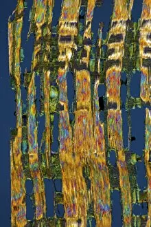
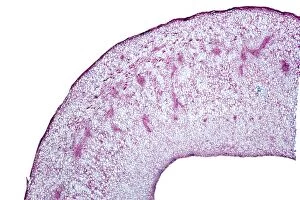
Structures Collection (#52)

"Captivating Structures from Around the World: A Visual Journey" Witness the mesmerizing sunrise behind the majestic Angkor Wat

Background imageStructures Collection: Asparagus stem tip, light micrograph

Asparagus stem tip, light micrograph
Asparagus stem tip. Light micrograph of a section through the tip of a narrow-leaved asparagus (Asparagus tenuifolius) stem

Background imageStructures Collection: Dendrobium orchid root, light micrograph

Dendrobium orchid root, light micrograph
Dendrobium orchid root. Light micrograph of a section through an aerial root from a Dendrobium sp. orchid. The outer tissue (velamen radicum, grey) is composed of hexagonal cells

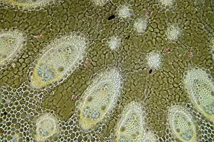
Background imageStructures Collection: Sharp rush stem, light micrograph

Sharp rush stem, light micrograph
Sharp rush stem. Light micrograph of a section through the stem of a sharp rush (Juncus acutus) plant. This arid-adapted plant (xerophyte) has scattered vascular bundles

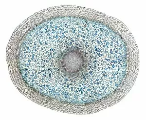
Background imageStructures Collection: Smilax root, light micrograph

Smilax root, light micrograph
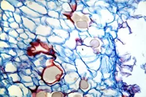
Smilax root. Light micrograph of a section through a root from a China root (Smilax china) plant. The cells are full of round starch grains (whitish-pink and blue)

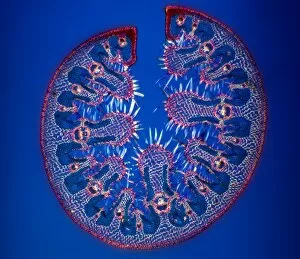
Background imageStructures Collection: Ammophila arenaria leaf, light micrograph

Ammophila arenaria leaf, light micrograph
Ammophila arenaria leaf. Polarised light micrograph of a section through a marram grass (Ammophila arenaria) leaf, showing the characteristics that help reduce water loss

Background imageStructures Collection: Date palm seed, light micrograph

Date palm seed, light micrograph
Date palm seed. Light micrograph of a section through a seed from a date palm (Phoenix dactylifera). Magnification: x37 when printed 10 centimetres wide

Background imageStructures Collection: Lily flower bud, light micrograph

Lily flower bud, light micrograph
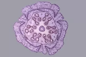
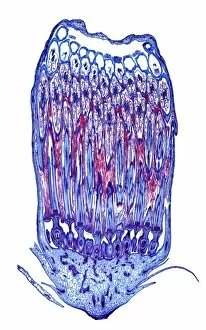
Lily flower bud. Light micrograph of a section through a young flower bud from a lily (Lilium sp.), showing pollen cells (circles) dividing within the anthers (inner ring)

Background imageStructures Collection: Celandine (Chelidonium majus) seeds

Celandine (Chelidonium majus) seeds
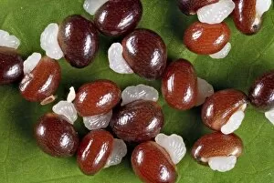
Greater celandine (Chelidonium majus) seeds. Close-up of seeds from the Greater celandine (Chelidonium majus) plant, showing elaiosomes (white)

Background imageStructures Collection: Female pine cone seed, light micrograph

Female pine cone seed, light micrograph
Female pine cone seed. Light micrograph of a section through a seed from a female pine (Pinus sp.) cone, showing the mature embryo and endosperm

Background imageStructures Collection: Beech tree leaf, light micrograph

Beech tree leaf, light micrograph
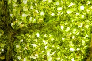
Beech tree leaf. Light micrograph of a section through the leaf of a common beech tree (Fagus sylvatica), showing the midrib

Background imageStructures Collection: Broad bean flower, light micrograph

Broad bean flower, light micrograph
Broad bean flower. Polarised light micrograph of a section through a flower from a broad bean (Vicia faba) plant, showing the stamens and stigma

Background imageStructures Collection: Pear rust fungus, light micrograph

Pear rust fungus, light micrograph
Pear rust fungus. Light micrograph of a section through tissue from a juniper tree infected with pear rust fungus (Gymnosporangium sabinae, lower right)

Background imageStructures Collection: Grey mould fungus, light micrograph

Grey mould fungus, light micrograph
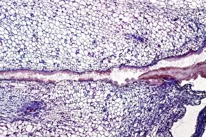
Grey mould fungus. Light micrograph of a section through onion tissue infected with grey mould fungus (Botrytis allii). Botrytis is a parasitic saprophyte mould that grows on wounded plants

Background imageStructures Collection: Corn smut infection, light micrograph

Corn smut infection, light micrograph
Corn smut infection. Light micrograph of a section through tissue from a corn (Zea mays) plant infected with corn smut fungus (Ustilago maydis)

Background imageStructures Collection: Bramble rust fungus spores

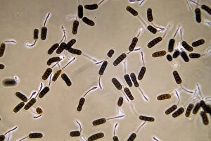
Bramble rust fungus spores. Light micrograph of teliospores from the bramble rust fungus (Phragmidium violaceum). Magnification: x37 when printed 10 centimetres wide

Background imageStructures Collection: Dahlia tuber, light micrograph

Dahlia tuber, light micrograph
Dahlia tuber. Polarised light micrograph of a section through the tuber of a Dahlia sp. plant, showing inulin crystals. The Dahlia produces root tubers that are mostly made up of scattered vascular

Background imageStructures Collection: Tomato fruit, light micrograph

Tomato fruit, light micrograph
Tomato fruit. Light micrograph of a section through the young fruit of a tomato (Solanum lycopersicum) plant, showing the seeds (ovules, oval) developing inside

Background imageStructures Collection: Horse-chestnut leaf, light micrograph

Horse-chestnut leaf, light micrograph
Horse-chestnut leaf. Light micrograph of a section through a leaf from a horse-chestnut, or conker, tree (Aesculus hippocastanum)

Background imageStructures Collection: Rose mildew fungus, light micrograph

Rose mildew fungus, light micrograph
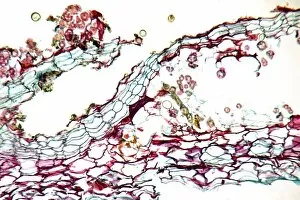
Rose mildew fungus. Light micrograph of a section through tissue from a rose plant infected with rose mildew fungus (Erysiphe pannosa)

Background imageStructures Collection: Pine root, light micrograph

Pine root, light micrograph
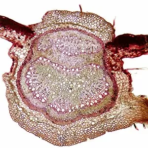
Pine root. Light micrograph of a section through the root of a pine (Pinus sp.) tree. From outer to inwards: outer layer of peridium (dark red); cortex - made up of parenchyma cells (red)
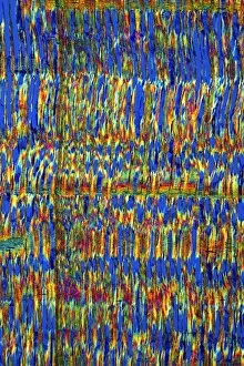
Background imageStructures Collection: Female pine cone scale, light micrograph

Female pine cone scale, light micrograph
Female pine cone scale. Light micrograph of a section through a scale from a young ovulate female pine (Pinus sp.) cone, bearing an ovule (upper centre)

Background imageStructures Collection: Pear scab infection, light micrograph

Pear scab infection, light micrograph
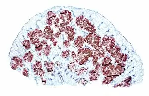
Pear scab infection. Light micrograph of a section through a pear (Pyrus sp.) infected with the Venturia pirinum fungus. This fungus causes scab, or black spot, of the pear fruit

Background imageStructures Collection: Black scab infection, light micrograph

Black scab infection, light micrograph
Black scab infection. Light micrograph of a section through a potato infected with black scab (Synchytrium endobioticum) fungus

Background imageStructures Collection: Lupin root nodules, light micrograph

Lupin root nodules, light micrograph
Lupin root nodules. Light micrograph of a section through the root nodule of a lupin (Lupinus lupin) plant. The plants root nodules are full of nitrogen-fixing Rhizobium sp

Background imageStructures Collection: Castor oil plant seed, light micrograph

Castor oil plant seed, light micrograph
Castor oil plant seed. Light micrograph of a section through the seed of a castor oil plant (Ricinus communis). The castor oil seed has an outer testa (not seen) which is poisonous

Background imageStructures Collection: Poppy flower bud, light micrograph

Poppy flower bud, light micrograph
Poppy flower bud. Light micrograph of a section through a flower bud from a poppy (Papaver sp.) plant. From outside to inside: calyx, made up of the sepals; corolla

Background imageStructures Collection: Dandelion flower, light micrograph

Dandelion flower, light micrograph
Dandelion flower. Light micrograph of a section through a flower from a dandelion (Taraxacum officinalis) plant. The flower stem (bottom) is made up of parenchyma cells (light blue)

Background imageStructures Collection: Common sundew, light micrograph

Common sundew, light micrograph
Common sundew. Light micrograph of a section through the leaf of a common sundew (Drosera rotundifolia) plant, showing the glandular hairs (dark)

Background imageStructures Collection: Great mullein hairs, light micrograph

Great mullein hairs, light micrograph
Great mullein hairs. Polarised light micrograph of hair-like structures, known as candelabra trichomes, from a great mullein (Verbascum thapsus) plant

Background imageStructures Collection: Powdery scab infection, light micrograph

Powdery scab infection, light micrograph
Powdery scab infection. Light micrograph of a section through a potato infected with powdery scab (Spongospora subterranea), showing pustules and spores. S

Background imageStructures Collection: Male pine cone, light micrograph

Male pine cone, light micrograph
Male pine cone. Light micrograph of a section through a male pine (Pinus sp.) cone, showing two microsporangia (pink) that contain numerous microspore pollen grains (blue)

Background imageStructures Collection: Sycamore leaf vein, light micrograph

Sycamore leaf vein, light micrograph
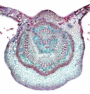
Sycamore leaf vein. Light micrograph of a section through the midrib (vein) of a leaf from a sycamore (Acer pseudoplatanus) tree

Background imageStructures Collection: Bean pod, light micrograph

Bean pod, light micrograph
Bean pod. Light micrograph of a section through the seed pod of a bean (Phaseolus sp.) plant. The pericarp (outer covering)

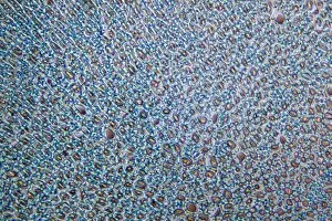
Background imageStructures Collection: African teak woody stem, light micrograph

African teak woody stem, light micrograph
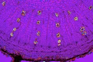
African teak woody stem. Polarised light micrograph of a section through the stem of an African teak (Pericopsis elata) plant, showing its diffuse-porous xylem tissue

Background imageStructures Collection: Venus flytrap leaf, light micrograph

Venus flytrap leaf, light micrograph
Venus flytrap leaf. Light micrograph of a section through the leaf of a Venus flytrap (Dionaea sp.), showing the digestive glands that line its interior

Background imageStructures Collection: Plum pocket infection, light micrograph

Plum pocket infection, light micrograph
Plum pocket infection. Light micrograph of a section through a plum gall caused by the plum pocket fungus (Taphrina pruni)

Background imageStructures Collection: Beech tree stem, light micrograph

Beech tree stem, light micrograph
Beech tree stem. Polarised light micrograph of a section through the woody stem of a common beech tree (Fagus sylvatica), showing diffuse-porous xylem vessels (dark blue)


For sale as Licensed Images

Choose your image, Select your licence and Download the media

"Captivating Structures from Around the World: A Visual Journey" Witness the mesmerizing sunrise behind the majestic Angkor Wat, a testament to Cambodia's rich history and architectural brilliance in Siem Reap. The grandeur of St Pauls Cathedral in Melbourne is amplified by its magnificent pipe organ, filling the air with enchanting melodies that resonate through time. Discover serenity as you behold a Buddha statue at Borobudur adorned with Dharmachakra Mudra hand position during a breathtaking sunrise in Magelang, Central Java. Gaze upon the stunning panorama of Victoria Harbour, Kowloon, and Hong Kong Island from atop Victoria Peak - an awe-inspiring structure showcasing China's vibrant cityscape. Experience an ethereal sunrise illuminating Dresden Frauenkirche, symbolizing resilience and rebirth after its reconstruction following World War II devastation in Germany. Pay homage to fallen heroes at Australian War Memorial in Canberra; this poignant structure stands as a reminder of sacrifice and valor for generations to come. Step into history on Kings Square (Market Place) Enfield Town, London; where centuries-old architecture meets bustling modernity around the iconic Kings Head Public House. Prepare to be amazed by Hollow-face illusion artwork that defies perception; these structures play tricks on our minds while captivating our imagination. Immerse yourself in Utrecht's charm as you witness a picturesque sunrise over Dom Tower and Vismarkt-Choorstraat along Oudegracht - an architectural marvel nestled within Netherlands' cultural heartland. Uncover ancient Roman artistry within painted murals and frescoes adorning rooms at Herculaneum ruins near Campania, Italy - preserving glimpses of past civilizations for eternity. Thurles Cathedral stands tall amidst Co Tipperary's lush landscapes in Ireland; explore its intricate design and bask in its spiritual ambiance.