
Tendon Collection (#8)
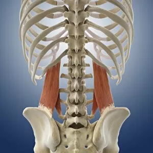
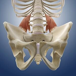
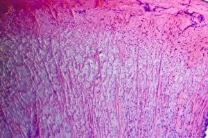
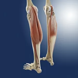
"Tendon: The Unsung Heroes of the Muscular System" The facial muscles of the human face, intricately labeled and interconnected


For sale as Licensed Images
Choose your image, Select your licence and Download the media
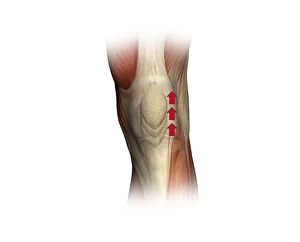
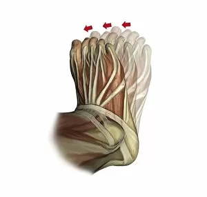
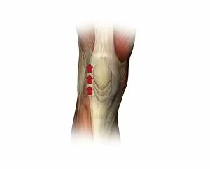
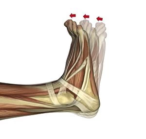
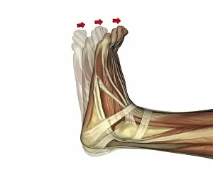
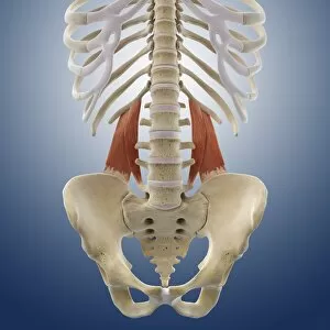
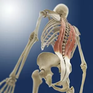
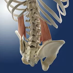
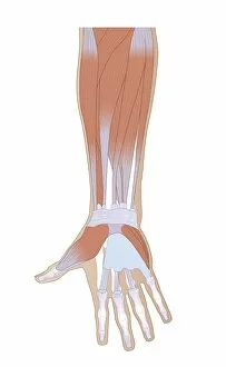
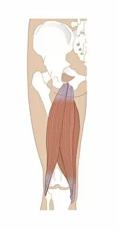
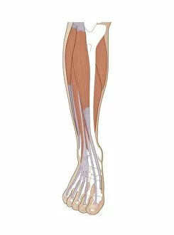
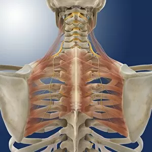
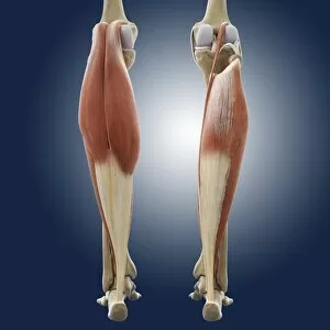
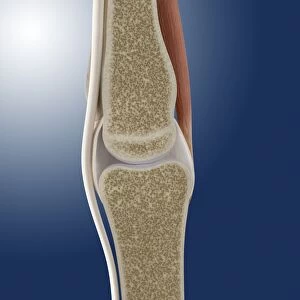
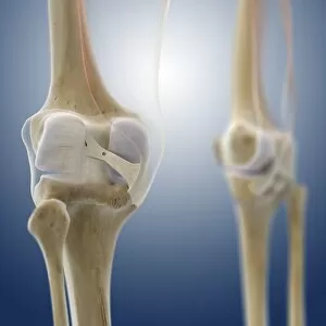
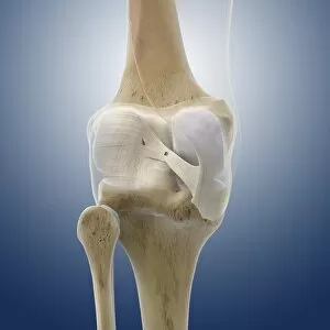
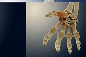
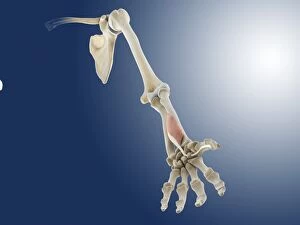
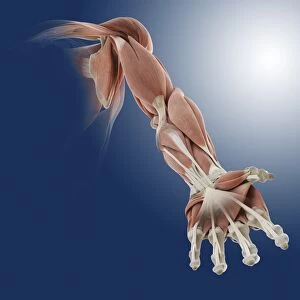
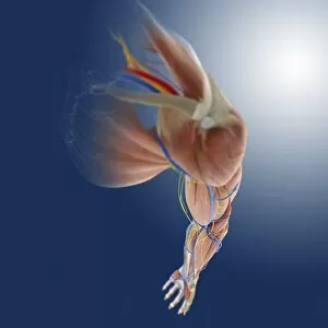
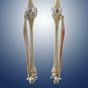
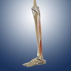
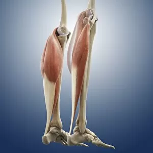
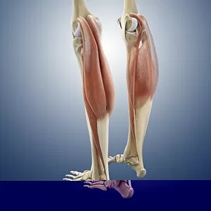
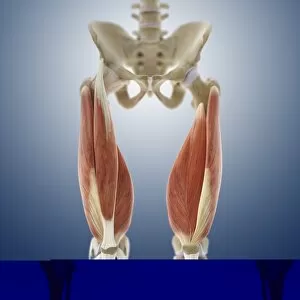
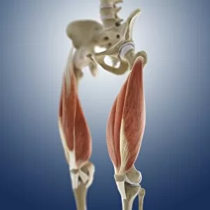
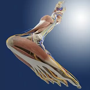
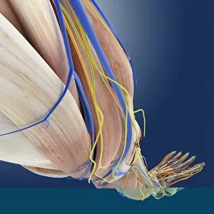
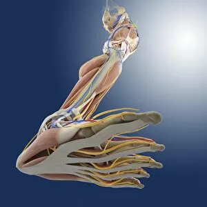
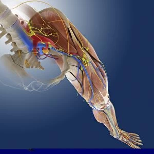
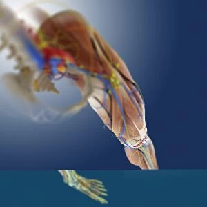
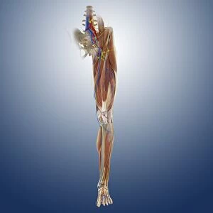
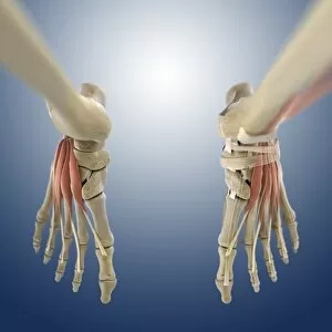
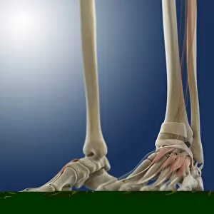
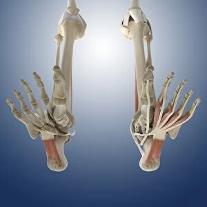
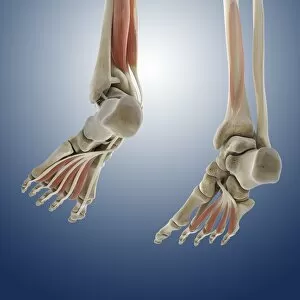
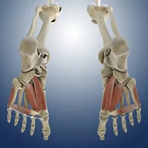
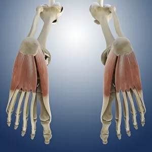
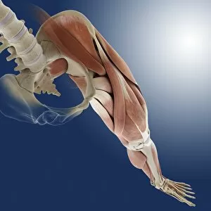
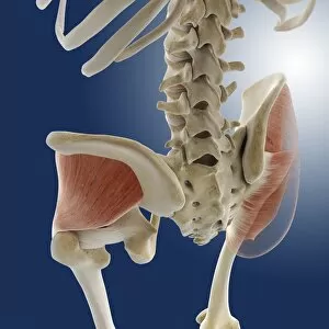
"Tendon: The Unsung Heroes of the Muscular System" The facial muscles of the human face, intricately labeled and interconnected, create a symphony of expressions that reflect our emotions. These muscles, part of the muscular system on our head, allow us to smile, frown, and convey countless other feelings. However, sometimes these they can become inflamed or irritated. Tendinitis of the shoulder is a painful condition that affects many individuals. An X-ray reveals this discomforting ailment in detail. Intriguingly captured in a line engraving from 19th-century France by an unknown artist, we witness the beauty and complexity anatomy. This illustration showcases how tendons connect muscles to bones with remarkable precision. Moving down to our feet - another masterpiece created by nature - a 19th-century illustration depicts foot anatomy in all its glory. It reveals not only skin and veins but also arteries, muscles, and bones working together harmoniously. Julien Bougle's colored plates superimposed on the human body provide an artistic representation showcasing various systems within us. Among them are tendons that play their crucial role silently yet efficiently. Delving deeper into internal structures brings us to an exquisite engraving depicting the intricate anatomy of organs. Here we see how tendons intertwine with other vital components like blood vessels and nerves to ensure proper functioning throughout our bodies. Zooming back out again to examine foot anatomy once more - this time showing skin texture alongside veins, arteries, muscles, and bones - we gain further appreciation for these unsung heroes called tendons. Our hands too rely heavily on healthy tendons for dexterity and strength as they perform daily tasks ranging from delicate movements to powerful grips. They enable us to write words like these or hold loved ones close with tender care. Yet even strong they can be vulnerable; an MRI image captures a ruptured Achilles tendon revealing just how crucial their integrity is for our mobility and overall well-being.