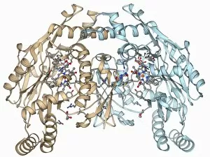
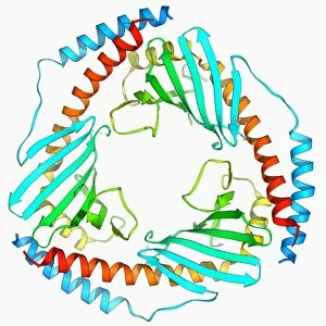
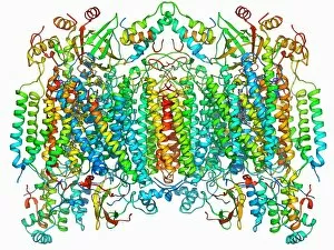
Tertiary Structure Collection (#7)

Tertiary structure is a fascinating aspect of molecular biology that plays a crucial role in the functionality and stability of various biological molecules

Background imageTertiary Structure Collection: Photosystem II molecule F006 / 9500

Photosystem II molecule F006 / 9500
Photosystem II. Molecular model of the photosystem II complex. Photosystems are protein complexes involved in photosynthesis

Background imageTertiary Structure Collection: Photosystem II molecule F006 / 9497

Photosystem II molecule F006 / 9497
Photosystem II. Molecular model of the photosystem II complex. Photosystems are protein complexes involved in photosynthesis

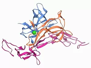
Background imageTertiary Structure Collection: Nerve growth factor bound to receptor F006 / 9501

Nerve growth factor bound to receptor F006 / 9501
Nerve growth factor bound to receptor. Molecular model of nerve growth factor (NGF) bound to the p75 neurotrophin receptor. NGF is a neurotrophin that acts on the development and function of nerves

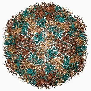
Background imageTertiary Structure Collection: Rhinovirus capsid, molecular model F006 / 9490

Rhinovirus capsid, molecular model F006 / 9490
Rhinovirus capsid, molecular model. This is human rhinovirus. The rhinovirus infects the upper respiratory tract and is the cause of the common cold. It is spread by coughs and sneezes

Background imageTertiary Structure Collection: H1 antigen from 1918 influenza virus F006 / 9495

H1 antigen from 1918 influenza virus F006 / 9495
H1 antigen from 1918 influenza virus

Background imageTertiary Structure Collection: HIV reverse transcription enzyme F006 / 9494

HIV reverse transcription enzyme F006 / 9494
HIV reverse transcription enzyme. Molecular model of the reverse transcriptase enzyme (blue and green) found in HIV (the human immunodeficiency virus)

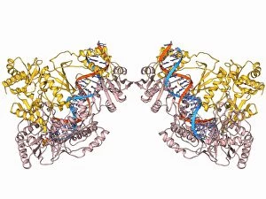
Background imageTertiary Structure Collection: EcoRV restriction enzyme molecule F006 / 9496

EcoRV restriction enzyme molecule F006 / 9496
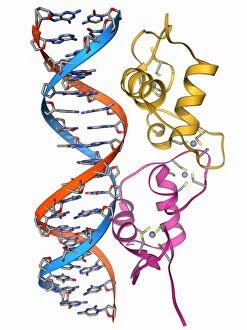
EcoRV restriction enzyme. Molecular model of the type II restriction enzyme EcoRV (pink and yellow) bound to a cleaved section of DNA (deoxyribonucleic acid, red and blue)

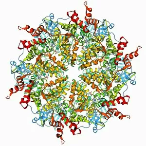
Background imageTertiary Structure Collection: Rubisco enzyme molecule F006 / 9491

Rubisco enzyme molecule F006 / 9491
Rubisco. Molecular model of the enzyme rubisco (ribulose bisphosphate carboxylase oxygenase). Rubisco is thought to be the most abundant and important protein found in nature

Background imageTertiary Structure Collection: Hammerhead ribozyme molecule F006 / 9492

Hammerhead ribozyme molecule F006 / 9492
Hammerhead ribozyme, molecular model. Ribozymes are RNA (ribonucleic acid) molecules that catalyse certain biochemical reactions

Background imageTertiary Structure Collection: Murine p97 protein molecule F006 / 9487

Murine p97 protein molecule F006 / 9487
Murine p97 protein, molecular model. This mouse enzyme, is involved in membrane fusion and ubiquitin-dependent protein degradation

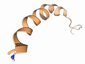
Background imageTertiary Structure Collection: Neuropeptide Y neurotransmitter molecule F006 / 9493

Neuropeptide Y neurotransmitter molecule F006 / 9493
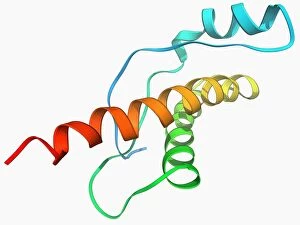
Neuropeptide Y neurotransmitter molecule. Molecular model showing the structure of the neurotransmitter neuropeptide Y (NPY). NPY is found in the brain and autonomic nervous system

Background imageTertiary Structure Collection: Interferon gamma molecule F006 / 9489

Interferon gamma molecule F006 / 9489
Interferon gamma, molecular model. Interferon gamma is produced by certain immune cells (T cells, dendritic cells and NK cells) as part of the immune response to invading pathogens and tumours

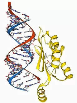
Background imageTertiary Structure Collection: RNA interference viral suppressor and RNA F006 / 9488

RNA interference viral suppressor and RNA F006 / 9488
RNA interference viral suppressor and RNA. Molecular model of the p19 protein (yellow) from a Tombusvirus, suppressing a double-stranded, small interfering RNA (siRNA) molecule (red and blue)

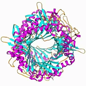
Background imageTertiary Structure Collection: Uricase enzyme, molecular model F006 / 9485

Uricase enzyme, molecular model F006 / 9485
Uricase, molecular model. This enzyme, also known as urate oxidase, catalyses the last stage of purine catabolism. It is absent from humans

Background imageTertiary Structure Collection: Yersinia pestis virulence factor F006 / 9486

Yersinia pestis virulence factor F006 / 9486
Yersinia pestis virulence factor. Molecular model of the V-antigen protein, a virulence factor in Yersinia pestis, the causative agent of the plague

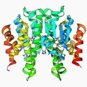
Background imageTertiary Structure Collection: Transcription factor and DNA molecule F006 / 9484

Transcription factor and DNA molecule F006 / 9484
Transcription factor and DNA molecule. Molecular model of glucocorticoid receptor (GR) transcription factor protein (pink and blue) complexed with a molecule of DNA (deoxyribonucleic acid)

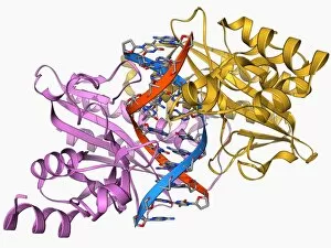
Background imageTertiary Structure Collection: Androgen receptor, molecular model F006 / 9483

Androgen receptor, molecular model F006 / 9483
Androgen receptor. Molecular model of the DNA-binding region of an androgen receptor (pink and yellow) complexed with DNA (deoxyribonucleic acid, blue and red)

Background imageTertiary Structure Collection: Human catalase, molecular model F006 / 9478

Human catalase, molecular model F006 / 9478
Human catalase, molecular model. This enzyme catalyses the break down of hydrogen peroxide to water and oxygen. Hydrogen peroxide is a highly toxic byproduct of a number of normal cellular processes

Background imageTertiary Structure Collection: Chinese scorpion toxin structure F006 / 9482

Chinese scorpion toxin structure F006 / 9482
Chinese scorpion toxin structure. Molecular model of the toxin BmBKTtx1, produced by the Chinese scorpion (Buthus martensi Karsch)
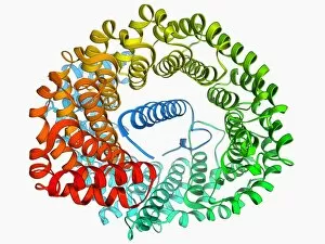
Background imageTertiary Structure Collection: Novel protein, molecular model F006 / 9481

Novel protein, molecular model F006 / 9481
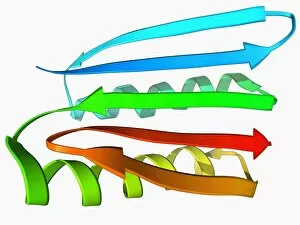
Novel protein. Molecular model of a computer-designed protein named TOP7

Background imageTertiary Structure Collection: Haemagglutinin protein subunit F006 / 9479

Haemagglutinin protein subunit F006 / 9479
Haemagglutinin protein subunit. Molecular model of the ectodomain of the haemagglutinin HA(2) subunit. Haemagglutinin is a surface protein from the influenza A virus

Background imageTertiary Structure Collection: Foot-and-mouth disease virus F006 / 9471

Foot-and-mouth disease virus F006 / 9471
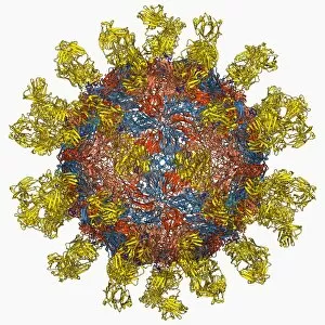
Foot-and-mouth disease virus. Molecular model of the foot-and-mouth disease (FMD) virus Aphtae epizooticae, with antibodies (immunoglobulins)

Background imageTertiary Structure Collection: RNA polymerase molecule F006 / 9475

RNA polymerase molecule F006 / 9475
RNA polymerase. Molecular model of RNA polymerase (beige) transcribing a strand of mRNA (messenger ribonucleic acid, pink) from a DNA (deoxyribonucleic acid) template (red and blue)

Background imageTertiary Structure Collection: Cytochrome c oxidase and antibody F006 / 9474

Cytochrome c oxidase and antibody F006 / 9474
Cytochrome c oxidase. Molecular model of a cytochrome c oxidase enzyme complexed with an antibody. Cytochrome molecules perform oxidation and reduction reactions for electron transport

Background imageTertiary Structure Collection: Human prion protein, molecular model F006 / 9477

Human prion protein, molecular model F006 / 9477
Human prion protein, molecular model. Prions are abnormal proteins that cause a group of fatal neurodegenerative diseases including BSE in cows and CJD in humans

Background imageTertiary Structure Collection: Herpesvirus immune inhibitor protein F006 / 9476

Herpesvirus immune inhibitor protein F006 / 9476
Herpesvirus immune inhibitor protein. Molecular model of the protein ICP47 from the herpes simplex virus-1 (HSV-1). This protein prevents antigens from the virus being presented to immune cells

Background imageTertiary Structure Collection: Poliovirus type 3 capsid, molecular model F006 / 9465

Poliovirus type 3 capsid, molecular model F006 / 9465
Poliovirus type 3 capsid, molecular model. This enterovirus causes poliomyelitis (polio) in humans, which affects the nervous system, sometimes leading to paralysis

Background imageTertiary Structure Collection: Importin heterodimer protein F006 / 9472

Importin heterodimer protein F006 / 9472
Importin heterodimer protein. Molecular model showing the importin heterodimer, a complex between importin alpha and importin beta. Importin imports other proteins into the cell nucleus

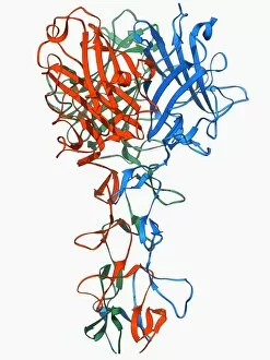
Background imageTertiary Structure Collection: Adenovirus fibre shaft protein F006 / 9473

Adenovirus fibre shaft protein F006 / 9473
Adenovirus fibre shaft protein, molecular model. This protein forms part of the shaft of the fibres or spikes by which adenoviruses attach to receptors on cells during the infection process

Background imageTertiary Structure Collection: Haemagglutinin viral surface protein F006 / 9470

Haemagglutinin viral surface protein F006 / 9470
Haemagglutinin viral surface protein. Molecular model of haemagglutinin, a surface protein from the influenza virus, complexed with a neutralising antibody

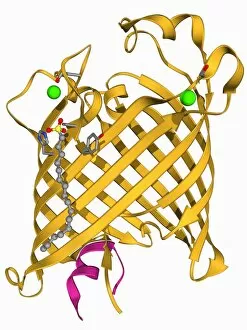
Background imageTertiary Structure Collection: Outer membrane phospholipase A molecule F006 / 9469

Outer membrane phospholipase A molecule F006 / 9469
Outer membrane phospholipase A. Molecular model of the integral membrane protein, outer membrane phospholipase A from the Escherichia coli bacterium

Background imageTertiary Structure Collection: Nickel-containing superoxide dismutase F006 / 9468

Nickel-containing superoxide dismutase F006 / 9468
Nickel-containing superoxide dismutase enzyme, molecular model. This enzyme scavenges and decomposes the potentially toxic first reduction product, superoxide, of aerobic respiration

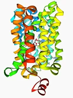
Background imageTertiary Structure Collection: Lactose transporter protein molecule F006 / 9466

Lactose transporter protein molecule F006 / 9466
Lactose transporter protein. Molecular model of the transmembrane transport protein lactose permease bound with a lactose homolog

Background imageTertiary Structure Collection: Aspirin drug target molecule F006 / 9464

Aspirin drug target molecule F006 / 9464
Aspirin drug target. Molecular model of the enzyme prostaglandin H2 synthase (PGHS), the target of the anti-inflammatory drug aspirin

Background imageTertiary Structure Collection: Archaeon enzyme, molecular model F006 / 9459

Archaeon enzyme, molecular model F006 / 9459
Archaeon enzyme. Molecular model of an enzyme from Thermoplasma acidophilum. This is the 20S proteasome. A proteasome is a complex type of proteinase (protein-digesting enzyme)

Background imageTertiary Structure Collection: Whooping cough toxin molecule F006 / 9463

Whooping cough toxin molecule F006 / 9463
Whooping cough toxin. Molecular model of the pertussis toxin from the bacterium Bordetella pertussis, the cause of whooping cough

Background imageTertiary Structure Collection: Photosynthetic reaction centre F006 / 9462

Photosynthetic reaction centre F006 / 9462
Photosynthetic reaction centre. Molecular model of the photosynthetic reaction centre from the purple bacterium Rhodopseudomonas viridis
Background imageTertiary Structure Collection: Dengue virus capsid, molecular model F006 / 9455

Dengue virus capsid, molecular model F006 / 9455
Dengue virus capsid, molecular model. This virus, transmitted by mosquito bites, causes the tropical disease dengue fever in humans

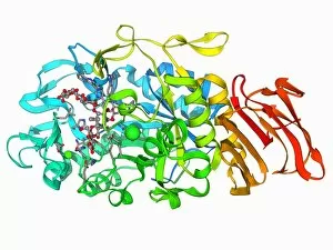
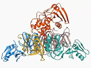
Background imageTertiary Structure Collection: Amylase, molecular model F006 / 9461

Amylase, molecular model F006 / 9461
Amylase. Molecular model of the enzyme alpha-amylase from the human pancreas. Amylase catalyses the breakdown of starch to sugars, and so starts the digestive process

Background imageTertiary Structure Collection: GST enzyme conferring DDT resistance F006 / 9460

GST enzyme conferring DDT resistance F006 / 9460
GST enzyme. Molecular model of a glutathione S-transferase (GST) enzyme from the malaria-carrying mosquito Anopheles gambiae

Background imageTertiary Structure Collection: Caspase-3 and inhibitor F006 / 9457

Caspase-3 and inhibitor F006 / 9457
Apopain protein and inhibitor. Molecular model of caspase-3, also known as apopain, complexed with an inhibitor. Caspase-3 is a protease, an enzyme that cleaves proteins

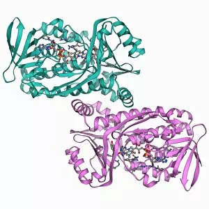
Background imageTertiary Structure Collection: Para-hydroxybenzoate hydroxylase molecule F006 / 9458

Para-hydroxybenzoate hydroxylase molecule F006 / 9458
Para-hydroxybenzoate hydroxylase, molecular model. This flavoprotein enzyme is involved in the degradation of aromatic compounds

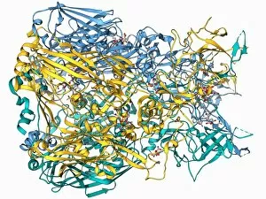
Background imageTertiary Structure Collection: Adenovirus hexon protein F006 / 9453

Adenovirus hexon protein F006 / 9453
Adenovirus hexon protein, molecular model. Hexon proteins are part of the protein coat or shell (capsid) of adenoviruses. In viruses

Background imageTertiary Structure Collection: Rous sarcoma virus capsid protein F006 / 9456

Rous sarcoma virus capsid protein F006 / 9456
Rous sarcoma virus capsid protein. Molecular model of the N-terminal domain of the Rous sarcoma virus capsid protein

Background imageTertiary Structure Collection: Nitric oxide synthase molecule F006 / 9452

Nitric oxide synthase molecule F006 / 9452
Nitric oxide synthase, molecular model. This enzyme catalyses the production of nitric oxide from L-arginine. Nitric oxide is involved in cellular signalling

Background imageTertiary Structure Collection: P32 mitochondrial matrix protein F006 / 9454

P32 mitochondrial matrix protein F006 / 9454
P32 mitochondrial matrix protein, molecular model. Also known as SF2-associated p32 (SF2P32), this protein is found in the matrix of cellular mitochondria

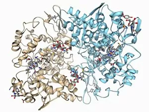
Background imageTertiary Structure Collection: Cytochrome c oxidase molecule F006 / 9447

Cytochrome c oxidase molecule F006 / 9447
Cytochrome c oxidase. Molecular model of a cytochrome c oxidase enzyme from the mitochondria of a cows heart. Cytochrome molecules perform oxidation and reduction reactions for electron transport

Background imageTertiary Structure Collection: ATPase and inhibitor, molecular model F006 / 9448

ATPase and inhibitor, molecular model F006 / 9448
ATPase and inhibitor. Computer model of an ATP synthase (ATPase) molecule from a mitochondrion complexed with its inhibitor protein IF1


For sale as Licensed Images

Choose your image, Select your licence and Download the media

Tertiary structure is a fascinating aspect of molecular biology that plays a crucial role in the functionality and stability of various biological molecules. One such example is the Manganese superoxide dismutase enzyme F006 / 9423, which possesses an intricate tertiary structure that allows it to efficiently scavenge harmful reactive oxygen species. Another intriguing molecule with a well-defined the Argonaute protein F006 / 9526. This protein acts as a key component of RNA-induced silencing complex F006 / 9586, enabling it to bind microRNAs and regulate gene expression through post-transcriptional mechanisms. The Immunoglobulin G antibody and egg white F006 / 9682 exhibit an interesting interaction where the tertiary structure of the antibody enables it to recognize specific antigens present in egg white, triggering an immune response. Cytochrome P450 complex F006 / 9669 showcases how multiple proteins come together to form a functional unit responsible for metabolizing drugs and toxins in our bodies. The precise arrangement of these proteins within their tertiary structures ensures efficient catalytic activity. Succinyl-CoA synthetase enzyme F006 / 9592 participates in the citric acid cycle (also known as Krebs cycle) by converting succinyl-CoA into ATP. Its unique tertiary structure facilitates substrate binding and enzymatic reactions essential for energy production. Viruses also possess captivating tertiary structures; one example being Foot-and-mouth disease virus F006 / 9556. Understanding its intricate capsid architecture aids researchers in developing effective vaccines against this highly contagious viral infection. Adenovirus penton base protein F006 / 9542 exhibits an elaborate tertiary structure critical for viral entry into host cells during infection. Studying this protein's conformation provides insights into potential therapeutic targets against adenoviral infections. Rhinovirus 16 capsid, molecular model F006/9431 represents another virus with a well-characterized tertiary structure.