Tibia Collection (#13)

"Tibia: Unveiling the Intricacies of the Human Knee Joint" Discover the fascinating world of tibia, a crucial bone in the human knee joint

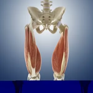
Background imageTibia Collection: Thigh muscles, artwork C013 / 4568

Thigh muscles, artwork C013 / 4568
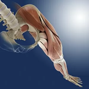
Thigh muscles. Computer artwork of an anterior view of some of the muscles and tendons (red and white) of the thighs and their attachment to the bones of the leg

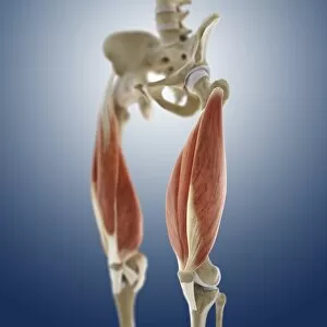
Background imageTibia Collection: Thigh muscles, artwork C013 / 4569

Thigh muscles, artwork C013 / 4569
Thigh muscles. Computer artwork of an oblique anterior view of some of the muscles and tendons (red and white) of the thighs and their attachment to the bones of the leg

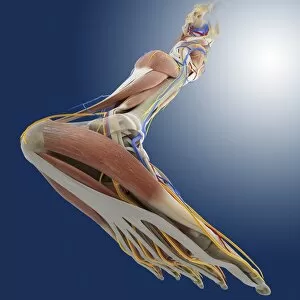
Background imageTibia Collection: Leg anatomy, artwork C013 / 4497

Leg anatomy, artwork C013 / 4497
Leg anatomy. Computer artwork of an oblique posterior view looking up a human leg, showing muscles and tendons (brown and white), bones and ligaments (white), arteries and veins (red and blue)

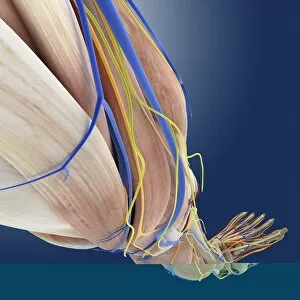
Background imageTibia Collection: Leg anatomy, artwork C013 / 4495

Leg anatomy, artwork C013 / 4495
Leg anatomy. Computer artwork of an oblique side view looking down a human leg, showing muscles and tendons (brown and white), bones and ligaments (white), arteries and veins (red and blue)

Background imageTibia Collection: Leg anatomy, artwork C013 / 4496

Leg anatomy, artwork C013 / 4496
Leg anatomy. Computer artwork of an oblique anterior view looking up a human leg, showing muscles and tendons (brown and white), bones and ligaments (white), arteries and veins (red and blue)

Background imageTibia Collection: Leg anatomy, artwork C013 / 4493

Leg anatomy, artwork C013 / 4493
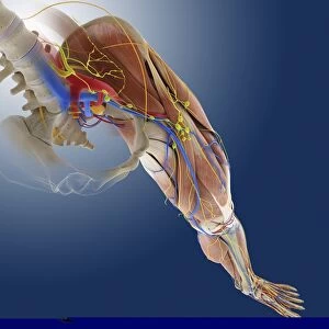
Leg anatomy. Computer artwork of an oblique anterior view looking down a human leg, showing muscles and tendons (brown and white), bones and ligaments (white), arteries and veins (red and blue)

Background imageTibia Collection: Leg anatomy, artwork C013 / 4494

Leg anatomy, artwork C013 / 4494
Leg anatomy. Computer artwork of an oblique anterior view looking down a human leg, showing muscles and tendons (brown and white), bones and ligaments (white), arteries and veins (red and blue)

Background imageTibia Collection: Leg ligaments, artwork

Leg ligaments, artwork
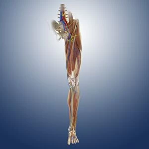
Leg anatomy. Computer artwork of an anterior view of a human leg, showing the muscles and tendons (brown and white), bones and ligaments (white), arteries and veins (red and blue), nerves (yellow)

Background imageTibia Collection: Foot muscles, artwork C013 / 4487

Foot muscles, artwork C013 / 4487
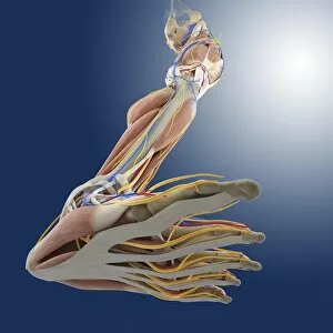
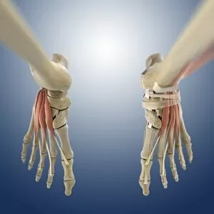
Foot muscles. Computer artwork of a view looking down on some of the muscles and tendons (red and white) of the upper surface of the foot

Background imageTibia Collection: Foot muscles, artwork C013 / 4486

Foot muscles, artwork C013 / 4486
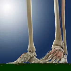
Foot muscles. Computer artwork of an oblique anterior view of some of the muscles and tendons (red and white) of the upper surface of the foot

Background imageTibia Collection: Foot muscles, artwork C013 / 4485

Foot muscles, artwork C013 / 4485
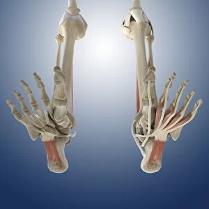
Foot muscles. Computer artwork of a view from below of some of the muscles and tendons (red and white) of the sole of the foot

Background imageTibia Collection: Foot muscles, artwork C013 / 4483

Foot muscles, artwork C013 / 4483
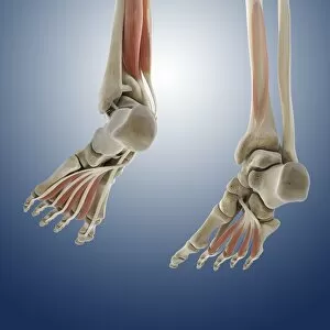
Foot muscles. Computer artwork of an oblique view from below and behind of some of the muscles and tendons (red and white) of the sole of the foot

Background imageTibia Collection: Outer ankle ligaments, artwork C013 / 4456

Outer ankle ligaments, artwork C013 / 4456
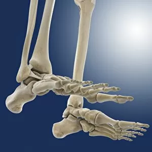
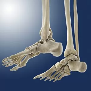
Outer ankle ligaments. Computer artwork of the bones and ligaments (white) of the feet and ankles from an oblique side view, with the outer side of the right foot at left

Background imageTibia Collection: Outer ankle ligaments, artwork C013 / 4455

Outer ankle ligaments, artwork C013 / 4455
Outer ankle ligaments. Computer artwork of the bones and ligaments (white) of the feet and ankles from an oblique side view, with the outer side of the left foot at upper centre

Background imageTibia Collection: Leg muscles, artwork C013 / 4429

Leg muscles, artwork C013 / 4429
Leg muscles. Computer artwork of the bones and outer muscles of the left leg, seen from the front (anterior view) at an oblique angle looking downwards

Background imageTibia Collection: Thigh hamstring muscles, artwork C013 / 4428

Thigh hamstring muscles, artwork C013 / 4428
Thigh hamstring muscles. Computer artwork of a posterior view of two of the three hamstring muscles (red) of the thighs. The muscles are the semimembranosus (inner) and semitendinosus (outer)

Background imageTibia Collection: Tibia insulae-chorab, Arabian tibia

Tibia insulae-chorab, Arabian tibia
Apair of Arabian tibia shells (Tibia insulae-chorab), This amazing, pointed gastropod can be found in the Indian Ocean in intertidal regions upto 50 metres deep

Background imageTibia Collection: Humerus of Achondroplastic Dwarf

Humerus of Achondroplastic Dwarf
Bones of Achondroplastic Dwarf. From the tomb of King Mersekha around the fourth Dynasty of Ancient Egypt

Background imageTibia Collection: Honeybee leg

Honeybee leg. Light micrograph of the hind leg of a honeybee (Apis mellifera). The tibia and first tarsal segment of the leg (large segments)

Background imageTibia Collection: Healthy knee, X-ray

Healthy knee, X-ray
Knee joint. Coloured X-ray of the healthy knee joint of an adult woman, seen from the side. The knee is the largest joint in the body

Background imageTibia Collection: Knee joint, X-ray

Knee joint, X-ray
Knee joint. Coloured X-ray of the healthy knee joint of a 15-year-old girl. The knee joint is composed of three bones; the femur (thigh bone, upper frame), the tibia (shin bone)

Background imageTibia Collection: Knee replacement, artwork

Knee replacement, artwork
Knee replacement. Artwork of a profile view of a prosthetic knee joint. The prostheses are implanted into the thigh bone (femur) and shinbone (tibia)

Background imageTibia Collection: Knee replacement, X-ray

Knee replacement, X-ray
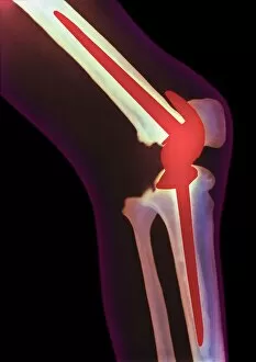
Knee replacement. Coloured X-ray of a side view of a prosthetic (artificial) knee joint (red) in the leg of a male patient

Background imageTibia Collection: Dislocated knee of baby, X-ray

Dislocated knee of baby, X-ray
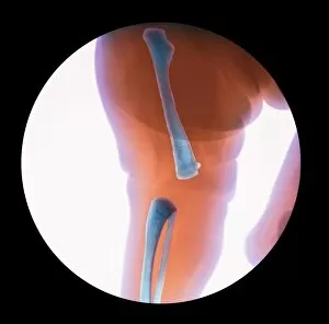
Congenital knee dislocation. Coloured X-ray of the dislocated and twisted knee of a female baby. The lower leg bones have twisted 180 degrees to the femur (at top)

Background imageTibia Collection: Coloured X-ray of fractured shin bone (tibia)

Coloured X-ray of fractured shin bone (tibia)
Broken leg. Coloured X-ray showing a fracture of the shin bone (tibia) of the leg. The tibia is the larger of the two bones of the lower leg, with the injury seen at centre right

Background imageTibia Collection: Kneecap fracture, X-ray

Kneecap fracture, X-ray
Kneecap fracture. Coloured X-ray of a broken kneecap (patella), as viewed from the side with the knee bent. The kneecap (far left, centre) is a disc of bone that covers the front of the knee joint

Background imageTibia Collection: Pinned ankle fracture, coloured X-ray

Pinned ankle fracture, coloured X-ray
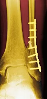
Pinned ankle fracture. Coloured X-ray of the ankle bones of a patient with a pinned ankle fracture. The malleolus (bony projection at the end of a bone) of the fibula (right)

Background imageTibia Collection: Knee meniscus tear

Knee meniscus tear
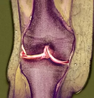
MODEL RELEASED. Knee meniscus tear. Coloured magnetic resonance imaging (MRI) scan of a coronal (front) section through the right knee joint of a 52 year old woman

Background imageTibia Collection: Coloured X-ray of a fractured tibia & fibula

Coloured X-ray of a fractured tibia & fibula
Pinned broken leg. Coloured X-ray of a pinned fracture of both the tibia and fibula (lower leg bones). The tibia (larger bone, centre) has been immobilised by metal pins (lower & upper centre)

Background imageTibia Collection: Corrected knee dislocation, X-ray

Corrected knee dislocation, X-ray
Corrected knee dislocation. Coloured X-ray of the dislocated and twisted knee of a female baby being manipulated back into place. The hand of the doctor is at bottom

Background imageTibia Collection: Pinned broken leg

Pinned broken leg. X-ray of a pinned fracture of both the tibia and fibula (lower leg bones). The tibia (larger bone, at centre) has been immobilised by metal pins (lower & upper centre)

Background imageTibia Collection: Broken leg

Broken leg. Coloured X-ray showing multiple fractures (centre) to the lower leg. Both the tibia (the thicker bone, foreground) and the fibula have been broken

Background imageTibia Collection: Artwork of rickets in tibia and fibula bones

Artwork of rickets in tibia and fibula bones
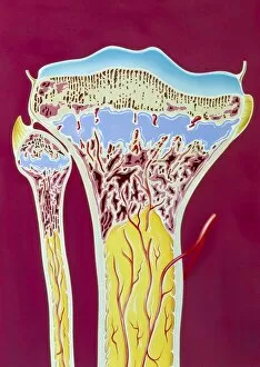
Rickets. Illustration of a cross section through the tibia (left) and fibula bones of the lower leg showing the signs of rickets - a bone disorder in which growing bones fail to mineralise properly

Background imageTibia Collection: Healthy ankle, X-ray

Healthy ankle, X-ray
Healthy ankle. Coloured X-ray of the ankle joint of an adult woman, in side view. The bones which form the ankle joint are the shin bone (tibia, at top) with the smaller fibula (behind it)

Background imageTibia Collection: Foot bones

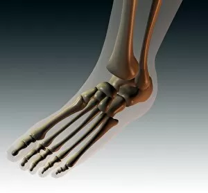
Foot bones, computer artwork. The toes are made up of the phalanx bones (phalanges), two for the big toe (lower left) and three for the others

Background imageTibia Collection: Knee joint from front and side, X-ray

Knee joint from front and side, X-ray
Knee joint from the front (left) and side (right), coloured X-rays. The knee is the largest joint in the human body. It is formed from the articulation of the thigh bone (femur)
Background imageTibia Collection: Artwork of human knee joint, side view

Artwork of human knee joint, side view

Background imageTibia Collection: Coloured X-ray of a human knee joint (front view)

Coloured X-ray of a human knee joint (front view)
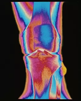
Knee joint. Coloured X-ray of a healthy human knee joint, in front view. Two bones meet at the knee, forming a joint that works like a hinge

Background imageTibia Collection: Coloured MRI scan of human knee joint, side view

Coloured MRI scan of human knee joint, side view
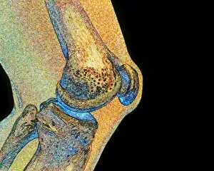
Knee joint. Coloured Magnetic Resonance Imaging (MRI) scan of a sagittal section through a human knee joint. Two bones (coloured blue) meet at the knee, forming a hinge-joint

Background imageTibia Collection: Coloured X-ray of a human knee joint (side view)

Coloured X-ray of a human knee joint (side view)
Knee joint. Coloured X-ray of a healthy human knee joint in side view, seen while bent. Two bones meet at the knee, forming a joint that works like a hinge

Background imageTibia Collection: Coloured X-ray image of a normal knee-joint

Coloured X-ray image of a normal knee-joint
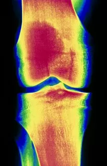
Normal knee. Coloured X-ray image of a healthy knee, showing the joint between the femur or thigh-bone (top) and tibia (the larger bone at bottom). This is a frontal view of the left leg

Background imageTibia Collection: Lower leg bones and ligaments

Lower leg bones and ligaments. Historical anatomical artwork of lower leg bones (yellow) and ligaments (pale blue). Ligaments are bands of fibrous tissue that hold bones together at their joints

Background imageTibia Collection: Arthritic knee

Arthritic knee
Arthritis. Computer artwork of an X-ray of an arthritic knee

Background imageTibia Collection: Healing ankle fracture, X-ray

Healing ankle fracture, X-ray
Healing ankle fracture, coloured X-rays. The ankle is seen from the front at left and from the side at right. The break is at the bottom of the fibula (left in front view)

Background imageTibia Collection: Knee effusion

Knee effusion. Coloured X-ray of the swelling of an injured knee caused by effusion. Effusion is a painful condition whereby fluid accumulates in the joint space, limiting movement

Background imageTibia Collection: Healed leg break, X-ray

Healed leg break, X-ray
Healed leg fracture. Coloured X-ray of a healed fracture of the tibia (shin bone) of an adult man. The two ends of the broken bone have healed causing some overgrowth of the bone at the fracture site


For sale as Licensed Images

Choose your image, Select your licence and Download the media

"Tibia: Unveiling the Intricacies of the Human Knee Joint" Discover the fascinating world of tibia, a crucial bone in the human knee joint. From normal foot X-rays to intricate diagrams of leg bones and hip structure, this caption takes you on an enlightening journey through various aspects related to tibia. Delve into the anatomy of a human knee joint with colored X-rays that showcase its complexity. Witness how knee joint prosthesis revolutionizes lives, as depicted by detailed X-ray images. Explore the fragility of damaged knee ligaments through captivating artwork, highlighting their vulnerability and importance for proper mobility. Uncover ancient myths as we delve into "The satyr Marsyas teaching Olympus to play, " where legends intertwine with our understanding of tibia's role in movement and strength. Marvel at medieval arms and armor, reminding us how vital strong limbs were throughout history. Witness fractures captured by ankle X-rays, emphasizing both resilience and vulnerability within our skeletal system. Immerse yourself in a comprehensive diagram showcasing bones, muscles, veins, arteries - every intricate detail that composes our remarkable human foot anatomy. Finally, bask in relief as healthy knees shine brightly through crystal-clear X-ray images. Appreciate these marvels that allow us to walk tall and explore life's wonders without hindrance. Join us on this captivating journey through visuals capturing tibia's significance within our bodies - from ancient tales to modern medical advancements. Let your curiosity be ignited as you uncover the secrets hidden beneath each image; embrace knowledge about one of nature's most incredible creations –the human body.