
Tibia Collection (#2)
"Tibia: Unveiling the Intricacies of the Human Knee Joint" Discover the fascinating world of tibia, a crucial bone in the human knee joint


For sale as Licensed Images
Choose your image, Select your licence and Download the media
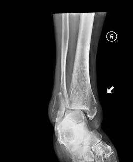
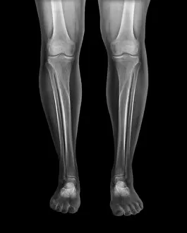
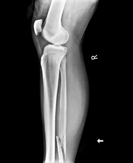
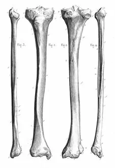
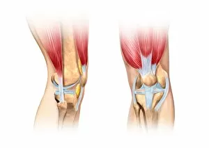
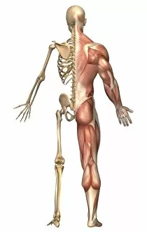
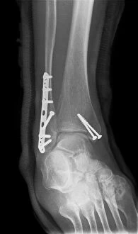
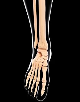
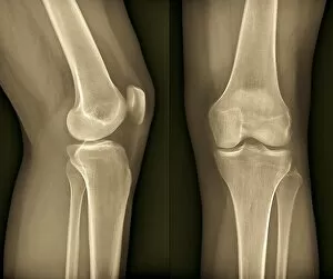
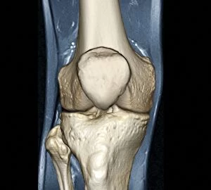
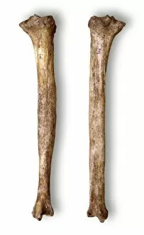
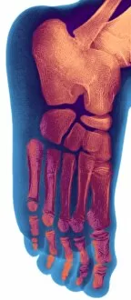
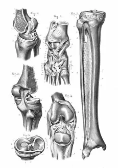
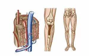
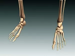
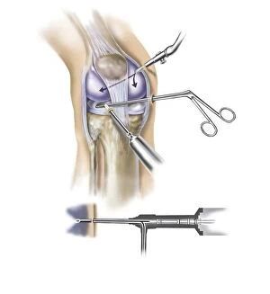
"Tibia: Unveiling the Intricacies of the Human Knee Joint" Discover the fascinating world of tibia, a crucial bone in the human knee joint. From normal foot X-rays to intricate diagrams of leg bones and hip structure, this caption takes you on an enlightening journey through various aspects related to tibia. Delve into the anatomy of a human knee joint with colored X-rays that showcase its complexity. Witness how knee joint prosthesis revolutionizes lives, as depicted by detailed X-ray images. Explore the fragility of damaged knee ligaments through captivating artwork, highlighting their vulnerability and importance for proper mobility. Uncover ancient myths as we delve into "The satyr Marsyas teaching Olympus to play, " where legends intertwine with our understanding of tibia's role in movement and strength. Marvel at medieval arms and armor, reminding us how vital strong limbs were throughout history. Witness fractures captured by ankle X-rays, emphasizing both resilience and vulnerability within our skeletal system. Immerse yourself in a comprehensive diagram showcasing bones, muscles, veins, arteries - every intricate detail that composes our remarkable human foot anatomy. Finally, bask in relief as healthy knees shine brightly through crystal-clear X-ray images. Appreciate these marvels that allow us to walk tall and explore life's wonders without hindrance. Join us on this captivating journey through visuals capturing tibia's significance within our bodies - from ancient tales to modern medical advancements. Let your curiosity be ignited as you uncover the secrets hidden beneath each image; embrace knowledge about one of nature's most incredible creations –the human body.