Tibia Collection (#3)

"Tibia: Unveiling the Intricacies of the Human Knee Joint" Discover the fascinating world of tibia, a crucial bone in the human knee joint

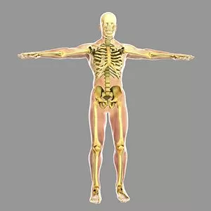
Background imageTibia Collection: Human skeletal system

Human skeletal system

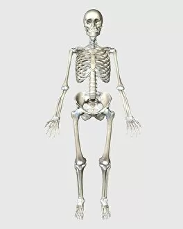
Background imageTibia Collection: Front view of human skeletal system

Front view of human skeletal system

Background imageTibia Collection: Woman doing gymnastics on the floor, with skeleton superimposed

Woman doing gymnastics on the floor, with skeleton superimposed

Background imageTibia Collection: Front view of human skeleton meditation

Front view of human skeleton meditation, isolated on white background

Background imageTibia Collection: 3D rendering of human skeletal system, rear view

3D rendering of human skeletal system, rear view

Background imageTibia Collection: Human male in athletic dynamic posture, with skeleton superimposed

Human male in athletic dynamic posture, with skeleton superimposed
Human male in athletic dynamic posture, with full skeleton superimposed

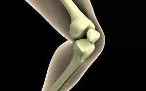
Background imageTibia Collection: Illustration of knee bone, bending

Illustration of knee bone, bending

Background imageTibia Collection: Anatomy of female body with organs

Anatomy of female body with organs

Background imageTibia Collection: X-ray image of knee

X-ray image of knee

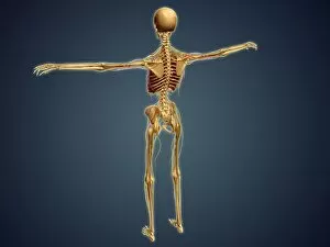
Background imageTibia Collection: Back view of human skeleton with nervous system, arteries and veins

Back view of human skeleton with nervous system, arteries and veins

Background imageTibia Collection: Conceptual image of human skeletal system

Conceptual image of human skeletal system

Background imageTibia Collection: Illustration of knee bone, straight

Illustration of knee bone, straight

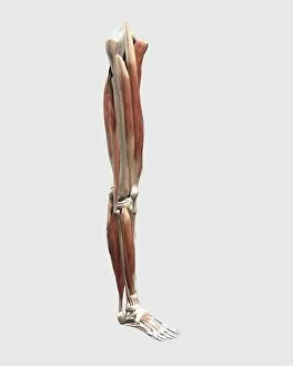
Background imageTibia Collection: Medical illustration of human leg muscles, bones and joints

Medical illustration of human leg muscles, bones and joints

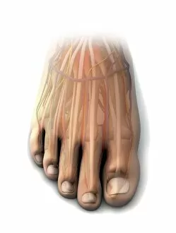
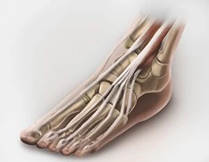
Background imageTibia Collection: Dorsal anatomy of the human foot

Dorsal anatomy of the human foot

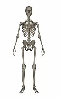
Background imageTibia Collection: Front view of human skeleton

Front view of human skeleton, isolated on white background

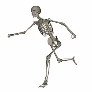
Background imageTibia Collection: Front view of human skeleton running

Front view of human skeleton running, isolated on white background

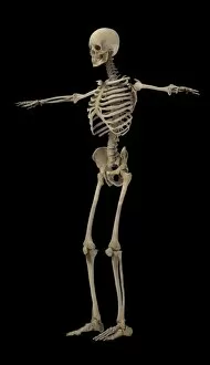
Background imageTibia Collection: 3D rendering of human skeletal system, side view

3D rendering of human skeletal system, side view

Background imageTibia Collection: Typical synovial joint

Typical synovial joint

Background imageTibia Collection: Detail of human knee showing arthroscopic surgical proceedures

Detail of human knee showing arthroscopic surgical proceedures

Background imageTibia Collection: Anterior compartment anatomy of left leg muscles and tendons

Anterior compartment anatomy of left leg muscles and tendons

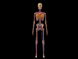
Background imageTibia Collection: Medical illustration of female skeleton with veins and arteries

Medical illustration of female skeleton with veins and arteries

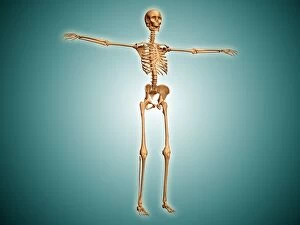
Background imageTibia Collection: Perspective view of human skeletal system

Perspective view of human skeletal system

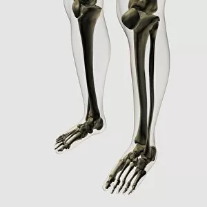
Background imageTibia Collection: Three dimensional view of human leg and feet bones

Three dimensional view of human leg and feet bones

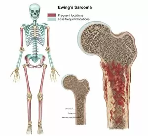
Background imageTibia Collection: Ewings sarcoma locations on the skeleton

Ewings sarcoma locations on the skeleton and detail of tumor on head of femur

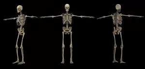
Background imageTibia Collection: 3D rendering of human skeletal system at different angles

3D rendering of human skeletal system at different angles

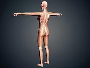
Background imageTibia Collection: Back view of female body with skeletal system superimposed

Back view of female body with skeletal system superimposed

Background imageTibia Collection: Medical illustration showing inflammation in human knee joint

Medical illustration showing inflammation in human knee joint
Medical illustration showing inflammation and pain in human knee joint, perspective view

Background imageTibia Collection: Cows, Anonymous, Herman van Swanevelt, 1636 - 1705

Cows, Anonymous, Herman van Swanevelt, 1636 - 1705

Background imageTibia Collection: Shepherd sits on a rock and plays the flute, a shepherdess spinning, goats and cows

Shepherd sits on a rock and plays the flute, a shepherdess spinning, goats and cows are around the couple, print maker: Jurriaan Cootwijck, Dating 1724 - 1798

Background imageTibia Collection: Trees Empire landscape with a well, Jan van de Velde (II), 1616

Trees Empire landscape with a well, Jan van de Velde (II), 1616

Background imageTibia Collection: Little boy blowing on a whistle, Theodore Schaepkens, 1825-1883

Little boy blowing on a whistle, Theodore Schaepkens, 1825-1883

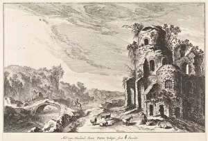
Background imageTibia Collection: Landscape with a bridge and a ruin, print maker: Pieter Nolpe, Adriaen van Nieulandt I

Landscape with a bridge and a ruin, print maker: Pieter Nolpe, Adriaen van Nieulandt I, Claes Jansz. Visscher II, 1623 - 1653

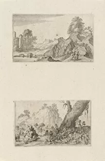
Background imageTibia Collection: Hikers in a hilly landscape and shepherd boy, Gillis van Scheyndel (I), 1605 - 1653

Hikers in a hilly landscape and shepherd boy, Gillis van Scheyndel (I), 1605 - 1653

Background imageTibia Collection: Couple making music, Jan van Somer, 1655 - 1700

Couple making music, Jan van Somer, 1655 - 1700

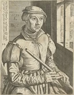
Background imageTibia Collection: The flautist, Bartholomeus Willemsz. Dolendo, Hendrick Hondius (I), 1589 - 1626

The flautist, Bartholomeus Willemsz. Dolendo, Hendrick Hondius (I), 1589 - 1626

Background imageTibia Collection: Flute Playing Shepherd and spinster, Nicolaes Pietersz. Berchem, 1652

Flute Playing Shepherd and spinster, Nicolaes Pietersz. Berchem, 1652

Background imageTibia Collection: Arcadian landscape with a shepherd near a well, Simon Klapmuts, 1770

Arcadian landscape with a shepherd near a well, Simon Klapmuts, 1770

Background imageTibia Collection: June, Jan van de Velde (II), 1616

June, Jan van de Velde (II), 1616

Background imageTibia Collection: In a hilly landscape is a shepherd playing the flute while a shepherdess lets her

In a hilly landscape is a shepherd playing the flute while a shepherdess lets her goats drink from a pond, in the background a house, print maker: Jan de Visscher, Dating 1643 - 1692

Background imageTibia Collection: Picture No. 10850679

Picture No. 10850679
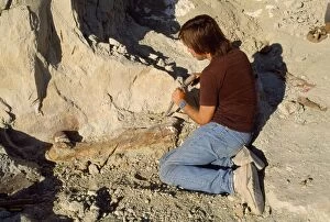
Dinosaur - excavating Allosaurus tibia. Morrison Formation, Jurassic. Date:

Background imageTibia Collection: Illustration of knee-joint

Illustration of knee-joint
Medicine: Human anatomy, knee-joint. Drawing

Background imageTibia Collection: Legs and feet of human skeleton, thigh bones, knees, lower leg bones, ankles, feet and toes

Legs and feet of human skeleton, thigh bones, knees, lower leg bones, ankles, feet and toes, front view

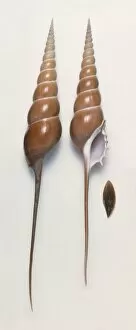
Background imageTibia Collection: Shinbone tibia (Tibia fusus) shells and operculum

Shinbone tibia (Tibia fusus) shells and operculum

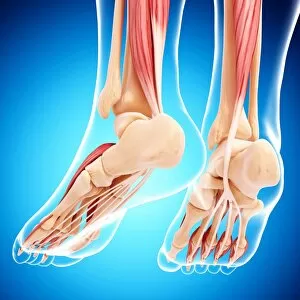
Background imageTibia Collection: Human foot musculature, artwork F007 / 1092

Human foot musculature, artwork F007 / 1092
Human foot musculature, computer artwork

Background imageTibia Collection: Human leg bones, artwork F007 / 4002

Human leg bones, artwork F007 / 4002
Human leg bones, computer artwork

Background imageTibia Collection: Human leg bones, artwork F007 / 4213

Human leg bones, artwork F007 / 4213
Human leg bones, computer artwork

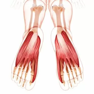
Background imageTibia Collection: Human foot musculature, artwork F007 / 4239

Human foot musculature, artwork F007 / 4239
Human foot musculature, computer artwork

Background imageTibia Collection: Human foot musculature, artwork F007 / 3948

Human foot musculature, artwork F007 / 3948
Human foot musculature, computer artwork


For sale as Licensed Images

Choose your image, Select your licence and Download the media

"Tibia: Unveiling the Intricacies of the Human Knee Joint" Discover the fascinating world of tibia, a crucial bone in the human knee joint. From normal foot X-rays to intricate diagrams of leg bones and hip structure, this caption takes you on an enlightening journey through various aspects related to tibia. Delve into the anatomy of a human knee joint with colored X-rays that showcase its complexity. Witness how knee joint prosthesis revolutionizes lives, as depicted by detailed X-ray images. Explore the fragility of damaged knee ligaments through captivating artwork, highlighting their vulnerability and importance for proper mobility. Uncover ancient myths as we delve into "The satyr Marsyas teaching Olympus to play, " where legends intertwine with our understanding of tibia's role in movement and strength. Marvel at medieval arms and armor, reminding us how vital strong limbs were throughout history. Witness fractures captured by ankle X-rays, emphasizing both resilience and vulnerability within our skeletal system. Immerse yourself in a comprehensive diagram showcasing bones, muscles, veins, arteries - every intricate detail that composes our remarkable human foot anatomy. Finally, bask in relief as healthy knees shine brightly through crystal-clear X-ray images. Appreciate these marvels that allow us to walk tall and explore life's wonders without hindrance. Join us on this captivating journey through visuals capturing tibia's significance within our bodies - from ancient tales to modern medical advancements. Let your curiosity be ignited as you uncover the secrets hidden beneath each image; embrace knowledge about one of nature's most incredible creations –the human body.