Transmission Electron Microscope Collection (#8)

The transmission electron microscope (TEM) has revolutionized our understanding of the intricate world within our bodies

Background imageTransmission Electron Microscope Collection: Kidney disease, TEM

Kidney disease, TEM
Kidney disease. Coloured transmission electron micrograph (TEM) of a section through the glomerulus of a kidney affected by membranous neuropathy

Background imageTransmission Electron Microscope Collection: Abnormal blood clot, TEM

Abnormal blood clot, TEM
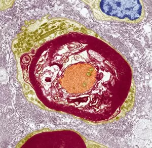
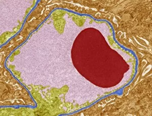
Abnormal blood clot. Coloured transmission electron micrograph (TEM) of a section through a blood vessel that is blocked by an abnormal blood clot (thrombus)

Background imageTransmission Electron Microscope Collection: Demyelinated nerve, TEM

Demyelinated nerve, TEM
Demyelinated nerve. Coloured transmission electron micrograph (TEM) of a section through an axon (a structure that transmits nerve impulses to other nerve cells) that has lost its myelin sheath

Background imageTransmission Electron Microscope Collection: Nerve demyelination, TEM

Nerve demyelination, TEM
Nerve demyelination. Coloured transmission electron micrograph (TEM) of a section through a Schwann cell and a nerve fibre, showing the early collapse of its myelin sheath

Background imageTransmission Electron Microscope Collection: Demyelinated nerve in multiple sclerosis

Demyelinated nerve in multiple sclerosis. Coloured transmission electron micrograph (TEM) of a section through an axon (a structure that transmits nerve impulses) that has lost its myelin sheath

Background imageTransmission Electron Microscope Collection: Alzheimers disease brain cell, TEM

Alzheimers disease brain cell, TEM
Alzheimers disease brain cell. Coloured transmission electron micrograph (TEM) of a neurofibrillary tangle in a nerve cell from the brain of a patient with Alzheimers disease

Background imageTransmission Electron Microscope Collection: Amyloidosis, TEM

Amyloidosis, TEM
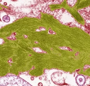
Amyloidosis. Coloured transmission electron micrograph (TEM) of a section through tissue with abnormal deposoits of amyloid protein (green)

Background imageTransmission Electron Microscope Collection: Hepatitis A virus particles, TEM

Hepatitis A virus particles, TEM
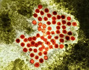
Hepatitis A virus. Coloured transmission electron micrograph (TEM) of a cluster of Hepatitis A virus particles, the cause of infectious hepatitis

Background imageTransmission Electron Microscope Collection: Influenzavirus B, TEM

Influenzavirus B, TEM
Influenzavirus B. Coloured transmission electron micrograph (TEM) of particles of the influenza B virus, which is the cause of influenza (flu) in humans

Background imageTransmission Electron Microscope Collection: SARS virus, TEM

SARS virus, TEM
SARS virus particle, coloured transmission electron micrograph (TEM). Severe acute respiratory syndrome (SARS) is an often fatal lung disease that first appeared in China in 2002

Background imageTransmission Electron Microscope Collection: Avian influenza virus, TEM

Avian influenza virus, TEM
Avian influenza virus, type A strain H5N1, coloured transmission electron micrograph (TEM). This virus was isolated in Vietnam, during the avian flu outbreak in early 2004

Background imageTransmission Electron Microscope Collection: Paramyxovirus, TEM

Paramyxovirus, TEM
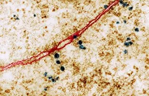
Paramyxovirus. Transmission electron micrograph (TEM) of paramyxovirus ribonuclear protein helices (red strands). These helices contain the viral genetic material, ribonucleic acid (RNA)

Background imageTransmission Electron Microscope Collection: Molluscum contagiosum virus, TEM

Molluscum contagiosum virus, TEM
Molluscum contagiosum virus. Coloured transmission electron micrograph (TEM) of two molluscum contagiosum virus (MCV) particles

Background imageTransmission Electron Microscope Collection: AIDS viruses budding from a cell, TEM

AIDS viruses budding from a cell, TEM
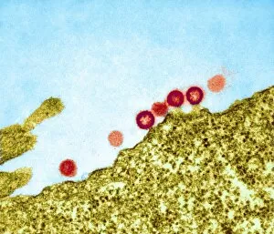
AIDS virus particles budding from the membrane of their host cell, coloured transmission electron micrograph (TEM). AIDS is a disease caused by the human immunodeficiency virus (HIV, red)

Background imageTransmission Electron Microscope Collection: Reovirus particles in a cell, TEM

Reovirus particles in a cell, TEM
Reovirus particles. Coloured transmission electron micrograph (TEM) of hundreds of particles (blue) of a reovirus infecting a host cell. The nucleus of the cell (green) is at centre

Background imageTransmission Electron Microscope Collection: AIDS virus particles, TEM

AIDS virus particles, TEM
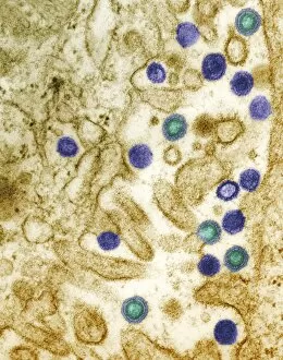
AIDS virus particles (blue and green) budding from the surface of a T4 lymphocyte cell, coloured transmission electron micrograph (TEM)

Background imageTransmission Electron Microscope Collection: Simian AIDS viruses, TEM

Simian AIDS viruses, TEM
Simian AIDS virus particles, coloured transmission electron micrograph (TEM). These virus particles (small, round) are bursting out of a cell (across bottom) after using the cell to replicate

Background imageTransmission Electron Microscope Collection: West Nile virus, TEM

West Nile virus, TEM
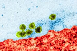
West Nile virus particles. Coloured transmission electron micrograph (TEM) of West Nile virus (WNV) particles (green) next to a host cell (red)

Background imageTransmission Electron Microscope Collection: Vaccinia viruses, TEM

Vaccinia viruses, TEM
Vaccinia virus particles. Coloured transmission electron micrograph (TEM) of sectioned vaccinia virus particles. The genetic cores (red) are covered by membrane layers (green)

Background imageTransmission Electron Microscope Collection: Pig retrovirus, TEM

Pig retrovirus, TEM
Pig retrovirus. Coloured transmission electron micrograph (TEM) of porcine endogenous retrovirus (PERV) in infected tissue. The retrovirus is the orange/red/blue structure at centre

Background imageTransmission Electron Microscope Collection: Programmed cell death

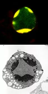
Programmed cell death. Comparison of a fluorescent light micrograph (top) and transmission electron micrograph (TEM, bottom) of programmed cell death (apoptosis)

Background imageTransmission Electron Microscope Collection: Golgi apparatus, TEM

Golgi apparatus, TEM
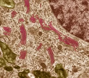
Golgi apparatus. Coloured transmission electron micrograph (TEM) of Golgi apparatus, also known as Golgi bodies, (pink) in a macrophage white blood cell

Background imageTransmission Electron Microscope Collection: Cell nucleolus, TEM

Cell nucleolus, TEM
Nucleolus. Coloured transmission electron micrograph of the nucleolus (blue), a component of the nucleus (orange) of a cell. The cell cytoplasm is green

Background imageTransmission Electron Microscope Collection: Cell organelles, TEM

Cell organelles, TEM
Cell organelles. Coloured transmission electron micrograph (TEM) of several organelles in an animal cell. At centre, the nucleus (dark purple, round) contains the cells genetic material (DNA)

Background imageTransmission Electron Microscope Collection: Ribosomes, TEM

Ribosomes, TEM
Ribosomes. Coloured transmission electron micrograph (TEM) of ribosomes (blue) passing through pores in a cells nuclear membrane (red)

Background imageTransmission Electron Microscope Collection: Lymph node, TEM

Lymph node, TEM
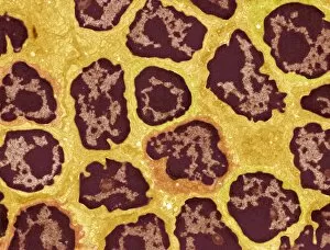
Lymph node. Coloured transmission electron micrograph (TEM) of a section through the peripheral corona of a lymph nodule, showing numerous small lymphocyte white blood cells (dark)

Background imageTransmission Electron Microscope Collection: Cell nucleus, TEM

Cell nucleus, TEM
Cell nucleus. Coloured transmission electron micrograph (TEM) of a section through a cell, showing the nucleus (large, spherical), and mitochondria (green)

Background imageTransmission Electron Microscope Collection: Glomerular capillary, TEM

Glomerular capillary, TEM
Glomerular capillary. Coloured transmission electron micrograph (TEM) of a section through kidney tissue, showing a capillary from a glomerulus. Glomeruli filter waste products from the blood

Background imageTransmission Electron Microscope Collection: Skin cell desmosomes, TEM

Skin cell desmosomes, TEM
Skin cell desmosomes. Coloured transmission electron micrograph (TEM) of a section through skin cells, showing numerous desmosomes (dark areas)

Background imageTransmission Electron Microscope Collection: Macrophage white blood cell, TEM

Macrophage white blood cell, TEM
Macrophage white blood cell. Coloured transmission electron micrograph (TEM) of a section through a macrophage from a lymph node, showing the nucleus (red) and numerous lysosomes (black)

Background imageTransmission Electron Microscope Collection: Pseudomonas sp. bacteria, TEM

Pseudomonas sp. bacteria, TEM
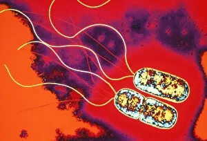
Pseudomonas sp. bacteria, coloured transmission electron micrograph (TEM). These are Gram- negative rod-shaped bacteria, with flagella (yellow strands) for movement

Background imageTransmission Electron Microscope Collection: Lactobacillus casei Shirota bacteria

Lactobacillus casei Shirota bacteria. Coloured transmission electron micrograph (TEM) of Lactobacillus casei Shirota, the strain of bacteria found in the Japanese probiotic product Yakult


For sale as Licensed Images

Choose your image, Select your licence and Download the media

The transmission electron microscope (TEM) has revolutionized our understanding of the intricate world within our bodies. With its high-resolution imaging capabilities, it allows us to delve deep into the microscopic realm and explore various cellular structures and processes. In the synapse nerve junction, TEM reveals a complex network of connections between neurons, highlighting their crucial role in transmitting signals throughout the nervous system. The rough endoplasmic reticulum, as seen through TEM, showcases its ribosome-studded surface responsible for protein synthesis. Witnessing a regenerating nerve cell under TEM provides insight into the remarkable ability of these cells to repair and restore function after injury. Meanwhile, observing a basophil white blood cell with this powerful microscope sheds light on its involvement in immune responses against allergens and parasites. TEM also unravels the fascinating process of myelination of nerve fibers - an essential mechanism for efficient signal conduction. Through detailed images captured by TEM, we can observe how myelin sheaths wrap around nerve fibers like protective insulation. Exploring further into nerve cells using TEM exposes their intricate structure comprising dendrites, axons, and synaptic terminals that enable communication within the nervous system. Additionally, mitochondria – often referred to as "the powerhouse of the cell" – are revealed in astonishing detail through TEM's lens. With precise imaging capabilities offered by TEM C014 / 1468, we can examine eye muscles at an unprecedented level. This aids in understanding their unique properties related to movement and coordination. Finally, studying Purkinje nerve cells with TEM C014 / 0583 allows us to appreciate their distinctive morphology within cerebellar tissue – characterized by elaborate branching patterns that contribute to motor coordination. In summary, transmission electron microscopy opens up a whole new dimension in biology research by providing unparalleled insights into cellular structures such as synapses, endoplasmic reticulum dynamics or regeneration processes while unraveling mysteries surrounding immune responses or myelination.