Transmission Electron Microscope Collection (#9)

The transmission electron microscope (TEM) has revolutionized our understanding of the intricate world within our bodies

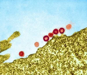
Background imageTransmission Electron Microscope Collection: AIDS viruses budding from a cell, TEM

AIDS viruses budding from a cell, TEM
AIDS virus particles budding from the membrane of their host cell, coloured transmission electron micrograph (TEM). AIDS is a disease caused by the human immunodeficiency virus (HIV, red)

Background imageTransmission Electron Microscope Collection: Reovirus particles in a cell, TEM

Reovirus particles in a cell, TEM
Reovirus particles. Coloured transmission electron micrograph (TEM) of hundreds of particles (blue) of a reovirus infecting a host cell. The nucleus of the cell (green) is at centre

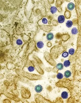
Background imageTransmission Electron Microscope Collection: AIDS virus particles, TEM

AIDS virus particles, TEM
AIDS virus particles (blue and green) budding from the surface of a T4 lymphocyte cell, coloured transmission electron micrograph (TEM)

Background imageTransmission Electron Microscope Collection: Simian AIDS viruses, TEM

Simian AIDS viruses, TEM
Simian AIDS virus particles, coloured transmission electron micrograph (TEM). These virus particles (small, round) are bursting out of a cell (across bottom) after using the cell to replicate

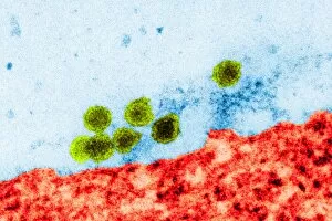
Background imageTransmission Electron Microscope Collection: West Nile virus, TEM

West Nile virus, TEM
West Nile virus particles. Coloured transmission electron micrograph (TEM) of West Nile virus (WNV) particles (green) next to a host cell (red)

Background imageTransmission Electron Microscope Collection: Vaccinia viruses, TEM

Vaccinia viruses, TEM
Vaccinia virus particles. Coloured transmission electron micrograph (TEM) of sectioned vaccinia virus particles. The genetic cores (red) are covered by membrane layers (green)

Background imageTransmission Electron Microscope Collection: Pig retrovirus, TEM

Pig retrovirus, TEM
Pig retrovirus. Coloured transmission electron micrograph (TEM) of porcine endogenous retrovirus (PERV) in infected tissue. The retrovirus is the orange/red/blue structure at centre

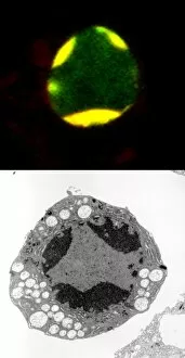
Background imageTransmission Electron Microscope Collection: Programmed cell death

Programmed cell death. Comparison of a fluorescent light micrograph (top) and transmission electron micrograph (TEM, bottom) of programmed cell death (apoptosis)

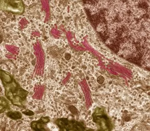
Background imageTransmission Electron Microscope Collection: Golgi apparatus, TEM

Golgi apparatus, TEM
Golgi apparatus. Coloured transmission electron micrograph (TEM) of Golgi apparatus, also known as Golgi bodies, (pink) in a macrophage white blood cell

Background imageTransmission Electron Microscope Collection: Cell nucleolus, TEM

Cell nucleolus, TEM
Nucleolus. Coloured transmission electron micrograph of the nucleolus (blue), a component of the nucleus (orange) of a cell. The cell cytoplasm is green

Background imageTransmission Electron Microscope Collection: Cell organelles, TEM

Cell organelles, TEM
Cell organelles. Coloured transmission electron micrograph (TEM) of several organelles in an animal cell. At centre, the nucleus (dark purple, round) contains the cells genetic material (DNA)

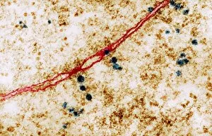
Background imageTransmission Electron Microscope Collection: Ribosomes, TEM

Ribosomes, TEM
Ribosomes. Coloured transmission electron micrograph (TEM) of ribosomes (blue) passing through pores in a cells nuclear membrane (red)

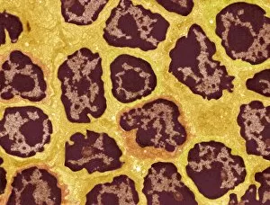
Background imageTransmission Electron Microscope Collection: Lymph node, TEM

Lymph node, TEM
Lymph node. Coloured transmission electron micrograph (TEM) of a section through the peripheral corona of a lymph nodule, showing numerous small lymphocyte white blood cells (dark)

Background imageTransmission Electron Microscope Collection: Cell nucleus, TEM

Cell nucleus, TEM
Cell nucleus. Coloured transmission electron micrograph (TEM) of a section through a cell, showing the nucleus (large, spherical), and mitochondria (green)

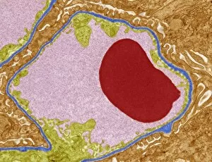
Background imageTransmission Electron Microscope Collection: Glomerular capillary, TEM

Glomerular capillary, TEM
Glomerular capillary. Coloured transmission electron micrograph (TEM) of a section through kidney tissue, showing a capillary from a glomerulus. Glomeruli filter waste products from the blood

Background imageTransmission Electron Microscope Collection: Skin cell desmosomes, TEM

Skin cell desmosomes, TEM
Skin cell desmosomes. Coloured transmission electron micrograph (TEM) of a section through skin cells, showing numerous desmosomes (dark areas)

Background imageTransmission Electron Microscope Collection: Macrophage white blood cell, TEM

Macrophage white blood cell, TEM
Macrophage white blood cell. Coloured transmission electron micrograph (TEM) of a section through a macrophage from a lymph node, showing the nucleus (red) and numerous lysosomes (black)

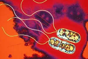
Background imageTransmission Electron Microscope Collection: Pseudomonas sp. bacteria, TEM

Pseudomonas sp. bacteria, TEM
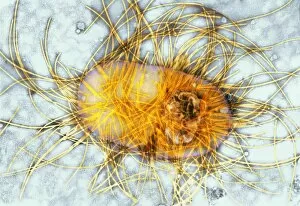
Pseudomonas sp. bacteria, coloured transmission electron micrograph (TEM). These are Gram- negative rod-shaped bacteria, with flagella (yellow strands) for movement

Background imageTransmission Electron Microscope Collection: Lactobacillus casei Shirota bacteria

Lactobacillus casei Shirota bacteria. Coloured transmission electron micrograph (TEM) of Lactobacillus casei Shirota, the strain of bacteria found in the Japanese probiotic product Yakult

Background imageTransmission Electron Microscope Collection: Mycobacterium bacteria, TEM

Mycobacterium bacteria, TEM
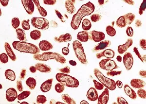
Mycobacterium bacteria. Coloured transmission electron micrograph (TEM) of Mycobacterium sp. bacteria. These are rod-shaped bacteria with a characteristic thick cell wall

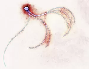
Background imageTransmission Electron Microscope Collection: Anaerobiospirillum thomasii flagella, TEM

Anaerobiospirillum thomasii flagella, TEM
Flagella of a spiral bacterium. Coloured transmission electron micrograph (TEM) of part of a bacterium with flagella attached (light blue)

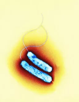
Background imageTransmission Electron Microscope Collection: Campylobacter oral bacteria, TEM

Campylobacter oral bacteria, TEM
Campylobacter bacteria. Coloured transmission electron micrograph (TEM) of two Campylobacter rectus bacteria. These Gram-negative rod-shaped bacteria have single flagellum (hair-like strand)

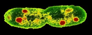
Background imageTransmission Electron Microscope Collection: Mycobacterium dividing, TEM

Mycobacterium dividing, TEM
Mycobacterium dividing. Coloured transmission electron micrograph (TEM) of a Mycobacterium sp. bacterium undergoing binary fission

Background imageTransmission Electron Microscope Collection: Chlamydia bacteria, TEM

Chlamydia bacteria, TEM
Chlamydia bacteria. Coloured transmission electron micrograph (TEM) of Chlamydia sp. bacteria (small blue spheres) in a human cell (large blue sphere)

Background imageTransmission Electron Microscope Collection: Erwinia bacteria, TEM

Erwinia bacteria, TEM
Erwinia bacteria. Coloured transmission electron micrograph (TEM) of the soil bacterium Erwinia carotovora carotovora. This gram-negative flagellated bacterium causes soft rot of plant crops such as

Background imageTransmission Electron Microscope Collection: Gut bacterium, TEM

Gut bacterium, TEM
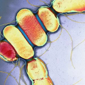
Gut bacterium. Coloured transmission electron micrograph (TEM) of a bacterium from the family Enterobacteriaceae. This family comprises Gram- negative rod-shaped bacteria

Background imageTransmission Electron Microscope Collection: Intestinal bacteria, TEM

Intestinal bacteria, TEM
Intestinal bacteria, coloured transmission electron micrograph (TEM). The TEM shows a section through the bacteria (red/yellow) and the wall of the intestines (bottom)

Background imageTransmission Electron Microscope Collection: Mycoplasma mycoides bacteria

Mycoplasma mycoides bacteria
Mycoplasma mycoides, coloured transmission electron micrograph (TEM). M. mycoides has a very small genome (total genetic material)

Background imageTransmission Electron Microscope Collection: Mycoplasma genitalium bacteria

Mycoplasma genitalium bacteria
Mycoplasma genitalium, coloured transmission electron micrograph (TEM). M. genitalium has the smallest genome (total genetic material) of all living organisms

Background imageTransmission Electron Microscope Collection: Stomatal complex, TEM

Stomatal complex, TEM
Stomal complex. Coloured transmission electron micrograph (TEM) of a stomatal (pore) complex in the young leaf of the pea plant (Pisum sativum)

Background imageTransmission Electron Microscope Collection: Gut bacterium reproducing, TEM

Gut bacterium reproducing, TEM
Gut bacterium reproducing. Coloured transmission electron micrograph (TEM) of a single bacterium dividing into two identical clone daughter bacteria

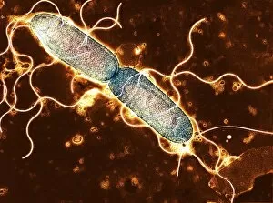
Background imageTransmission Electron Microscope Collection: Salmonella, TEM

Salmonella, TEM
Salmonella. Coloured transmission electron micrograph (TEM) of Salmonella paratyphi B bacteria (also called Salmonella schotmulleri)

Background imageTransmission Electron Microscope Collection: Q fever bacteria, Coxiella burnetii, TEM

Q fever bacteria, Coxiella burnetii, TEM
Q fever bacteria (Coxiella burnetii), coloured transmission electron micrograph (TEM). Q fever is a disease present in livestock that can be spread to humans by inhalation of contaminated particles

Background imageTransmission Electron Microscope Collection: Gonorrhoea bacteria, TEM

Gonorrhoea bacteria, TEM
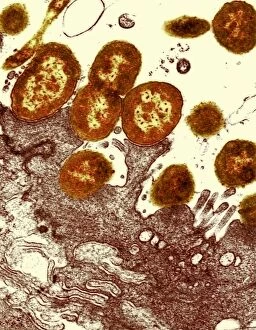
Gonorrhoea bacteria. Coloured transmission electron micrograph (TEM) of a Neisseria gonorrhoeae bacteria diplococci (pair of cells, light orange) infecting a human epithelial cell (dark orange)

Background imageTransmission Electron Microscope Collection: Pigment molecule, TEM

Pigment molecule, TEM
Pigment molecule, coloured transmission electron micrograph (TEM). This is a molecule of a pigment protein called alpha-crustacyanin

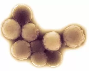
Background imageTransmission Electron Microscope Collection: MRSA bacteria

MRSA bacteria. Coloured transmission electron micrograph (TEM) of Methicillin-resistant Staphylococcus aureus (MRSA), a Gram-positive, round (coccus) bacterium

Background imageTransmission Electron Microscope Collection: MRSA bacteria, TEM

MRSA bacteria, TEM
MRSA bacteria. Coloured transmission electron micrograph (TEM) of methicillin-resistant Staphylococcus aureus (MRSA), a gram-positive, round (coccus) bacterium

Background imageTransmission Electron Microscope Collection: Streptococcus oral bacteria, TEM

Streptococcus oral bacteria, TEM
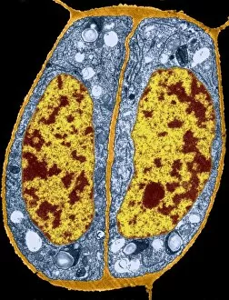
Streptococcus oral bacteria. Coloured transmission electron micrograph (TEM) of two Streptococcus sanguinis bacteria. They are undergoing nuclear division (mitosis)

Background imageTransmission Electron Microscope Collection: Poliovirus particles, TEM

Poliovirus particles, TEM
Poliovirus particles. Coloured transmissionelectron micrograph (TEM) of poliovirus particles.Each virus consists of a protein coat (capsid)surrounding a core of RNA (ribonucleicacid)


For sale as Licensed Images

Choose your image, Select your licence and Download the media

The transmission electron microscope (TEM) has revolutionized our understanding of the intricate world within our bodies. With its high-resolution imaging capabilities, it allows us to delve deep into the microscopic realm and explore various cellular structures and processes. In the synapse nerve junction, TEM reveals a complex network of connections between neurons, highlighting their crucial role in transmitting signals throughout the nervous system. The rough endoplasmic reticulum, as seen through TEM, showcases its ribosome-studded surface responsible for protein synthesis. Witnessing a regenerating nerve cell under TEM provides insight into the remarkable ability of these cells to repair and restore function after injury. Meanwhile, observing a basophil white blood cell with this powerful microscope sheds light on its involvement in immune responses against allergens and parasites. TEM also unravels the fascinating process of myelination of nerve fibers - an essential mechanism for efficient signal conduction. Through detailed images captured by TEM, we can observe how myelin sheaths wrap around nerve fibers like protective insulation. Exploring further into nerve cells using TEM exposes their intricate structure comprising dendrites, axons, and synaptic terminals that enable communication within the nervous system. Additionally, mitochondria – often referred to as "the powerhouse of the cell" – are revealed in astonishing detail through TEM's lens. With precise imaging capabilities offered by TEM C014 / 1468, we can examine eye muscles at an unprecedented level. This aids in understanding their unique properties related to movement and coordination. Finally, studying Purkinje nerve cells with TEM C014 / 0583 allows us to appreciate their distinctive morphology within cerebellar tissue – characterized by elaborate branching patterns that contribute to motor coordination. In summary, transmission electron microscopy opens up a whole new dimension in biology research by providing unparalleled insights into cellular structures such as synapses, endoplasmic reticulum dynamics or regeneration processes while unraveling mysteries surrounding immune responses or myelination.