White Blood Cell Collection (#12)

"Guardians of Health: Exploring the Mighty White Blood Cell" T lymphocytes and cancer cells: Unveiling the battle within, as T lymphocytes wage war against cancer cells

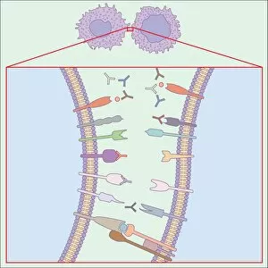
Background imageWhite Blood Cell Collection: Immune system, artwork

Immune system, artwork
Immune system. Computer artwork showing how T-cells (right), antibodies (Y-shaped) and antigen-presenting cells (APC, left) interact during an immune response

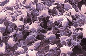
Background imageWhite Blood Cell Collection: Liver macrophage cells, SEM

Liver macrophage cells, SEM
Liver macrophage cells. Coloured scanning electron micrograph (SEM) of liver macrophages, also known as Kupffer cells. Macrophages are a type of large white blood cell that recognise foreign

Background imageWhite Blood Cell Collection: Human immune response, artwork

Human immune response, artwork
Human immune response. Computer artwork showing the interactions of the various white blood cells (purple) involved during an immune response in the human body

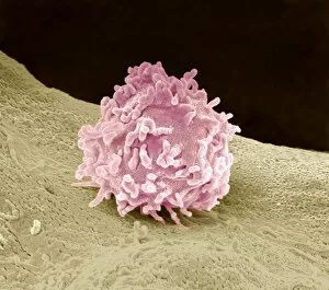
Background imageWhite Blood Cell Collection: Macrophage cell, SEM

Macrophage cell, SEM
Macrophage cell. Coloured scanning electron micrograph (SEM) of a macrophage white blood cell. Macrophages are cells of the bodys immune system

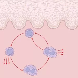
Background imageWhite Blood Cell Collection: Immune response to chronic inflammation

Immune response to chronic inflammation. Computer artwork showing the interaction of T cell, lymphocyte, and macrophage white blood cells in response to chronic inflammation of the skin (top)

Background imageWhite Blood Cell Collection: Skin damage immune response, artwork

Skin damage immune response, artwork
Skin damage immune response. Computer artwork showing the bodys immune response to pathogens (red dots) entering through a skin wound (upper centre)

Background imageWhite Blood Cell Collection: Activated platelets, SEM

Activated platelets, SEM
Activated platelets. Coloured scanning electron micrograph (SEM) of activated platelets, or thrombocytes, starting to adhere to exposed sub-endothelium on the inner surface of a vein

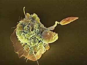
Background imageWhite Blood Cell Collection: Neutrophil engulfing fungus, SEM

Neutrophil engulfing fungus, SEM
Neutrophil engulfing fungus. Coloured scanning electron micrograph (SEM) of a neutrophil white blood cell (orange) engulfing and destroying a Candida albicans (thrush) fungal hypha (green)

Background imageWhite Blood Cell Collection: Liver macrophage cell, SEM

Liver macrophage cell, SEM
Liver macrophage cell. Coloured scanning electron micrograph (SEM) of a liver macrophage, also known as a Kupffer cell. Macrophages are a type of large white blood cell that recognise foreign

Background imageWhite Blood Cell Collection: Macrophage attacking a foreign body, SEM

Macrophage attacking a foreign body, SEM
Macrophage attacking a foreign body. Coloured scanning electron micrograph (SEM) of a macrophage white blood cell (round, left) engulfing a Leishmania mexicana protozoan parasite (long, right)

Background imageWhite Blood Cell Collection: Granulocyte, SEM

Granulocyte, SEM
Granulocyte. Coloured scanning electron micrograph (SEM) of a granulocyte in bone marrow. Granulocytes are a category of white blood cell characterized by the presence of granules in their cytoplasm

Background imageWhite Blood Cell Collection: Blood cells, artwork

Blood cells, artwork
Blood cells. Cut-away artwork of a blood vessel showing the blood cells inside. The majority of blood cells are erythrocytes (red blood cells)

Background imageWhite Blood Cell Collection: Granulocytes, SEM

Granulocytes, SEM
Granulocytes. Coloured scanning electron micrograph (SEM) of granulocytes in bone marrow. Granulocytes are a category of white blood cell characterized by the presence of granules in their cytoplasm

Background imageWhite Blood Cell Collection: Woundcare gel therapy, artwork

Woundcare gel therapy, artwork
Woundcare gel therapy. Artwork showing the cellular effects of woundcare therapy using a platelet-leucocyte enriched gel to support soft tissue and bone healing

Background imageWhite Blood Cell Collection: Platelet activation, artwork

Platelet activation, artwork
actication of PRP gel

Background imageWhite Blood Cell Collection: Frog blood, light micrograph

Frog blood, light micrograph. Unlike mammalian erythrocytes (red blood cells) mature frog erythrocytes retain their nuclei (black dots). Also seen are a few white blood cells (brown dots)
Background imageWhite Blood Cell Collection: Fish blood, light micrograph

Fish blood, light micrograph. Unlike mammalian erythrocytes (red blood cells) mature fish erythrocytes retain their nuclei (black dots). Also seen are a few white blood cells (brown dots)

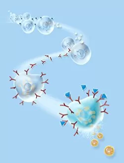
Background imageWhite Blood Cell Collection: Allergic response, artwork

Allergic response, artwork
Allergic response. Computer artwork showing the process that produces an allergic reaction. Antigens (blue), such as pollen

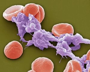
Background imageWhite Blood Cell Collection: Red blood cells and platelets, SEM

Red blood cells and platelets, SEM
Red blood cells and platelets. Coloured scanning electon micrograph (SEM) of human erythrocytes (red blood cells) and a platelet aggregate (purple)

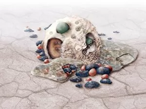
Background imageWhite Blood Cell Collection: Macrophage engulfing pathogen, artwork

Macrophage engulfing pathogen, artwork
Macrophage engulfing pathogen. Artwork of a macrophage cell (pink) engulfing a pathogen (green), a process known as phagocytosis. The pathogen will be destroyed once it has been engulfed

Background imageWhite Blood Cell Collection: Macrophage engulfing pathogens, artwork

Macrophage engulfing pathogens, artwork
Macrophage engulfing pathogens. Artwork of a macrophage cell (pink) engulfing pathogens (green), a process known as phagocytosis. The pathogens will be destroyed once they have been engulfed

Background imageWhite Blood Cell Collection: Malaria infection, artwork

Malaria infection, artwork
Malaria infection. Computer artwork of a macrophage white blood cell (right) extending pseudopodia towards a red blood cell (left) infected with the Plasmodium fulciparum malaria parasite

Background imageWhite Blood Cell Collection: Blood cells, SEM

Blood cells, SEM
Blood cells. Coloured scanning electron micrograph (SEM) of a human red blood cell (erythrocyte, red) and a white blood cell (leucocyte, blue). These are the two main cell components of blood

Background imageWhite Blood Cell Collection: Macrophage cells, TEM

Macrophage cells, TEM
Macrophage cells, coloured transmission electron micrograph (TEM). The cell nuclei are purple. Mitochondria (dark pink ovals) in the cytoplasm produce energy for the cell

Background imageWhite Blood Cell Collection: HIV particles exiting a cell, artwork

HIV particles exiting a cell, artwork
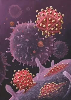
HIV particles exiting a cell. Artwork of HIV (human immunodeficiency virus) particles exiting a T-lymphocyte (white blood cell). HIV causes AIDS (acquired immune deficiency syndrome)

Background imageWhite Blood Cell Collection: Spleen cell, TEM

Spleen cell, TEM
Spleen cell. Coloured transmission electron micrograph (TEM) of a section through a cell in the spleen, an organ located in the upper left side of the abdomen in vertebrate animals

Background imageWhite Blood Cell Collection: Spleen cells, TEM

Spleen cells, TEM
Spleen cells. Coloured transmission electron micrograph (TEM) of a section through cells in the spleen, an organ located in the upper left side of the abdomen in vertebrate animals

Background imageWhite Blood Cell Collection: Lung alveoli and blood cells, TEM

Lung alveoli and blood cells, TEM
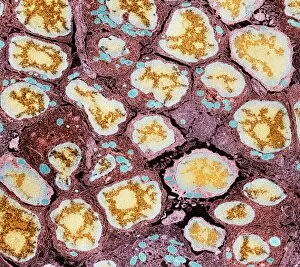
Lung alveoli and blood cells. Coloured transmission electron micrograph (TEM) of a section through lung alveoli and blood cells


For sale as Licensed Images

Choose your image, Select your licence and Download the media

"Guardians of Health: Exploring the Mighty White Blood Cell" T lymphocytes and cancer cells: Unveiling the battle within, as T lymphocytes wage war against cancer cells, SEM C001 / 1679. Neutrophil engulfing MRSA: Witness the incredible defense mechanism as a neutrophil engulfs MRSA bacteria, SEM C018 / 8596. Dendritic cells artwork: Unlocking the secrets of immune response with stunning artwork depicting dendritic cells at work. TEM of human white blood cell bearing HLA antigen: Peering into the intricate world of immunity through a transmission electron microscope image showcasing a white blood cell displaying HLA antigens. Blood cells in harmony: A mesmerizing glimpse into our life force - an ensemble of diverse blood cells working together for our well-being. Coloured SEM of a white blood cell (lymphocyte): Dive deep into the vibrant realm of lymphocytes captured in this captivating colored scanning electron microscope image. Basophil white blood cell: Discovering the lesser-known heroes among us - basophilic white blood cells that play crucial roles in allergic reactions and inflammation. Bacteria infecting a macrophage, SEM: Witness how macrophages confront invading bacteria head-on under high-resolution scanning electron microscopy imagery. Blood coagulation cascade artwork: Unraveling the intricacies behind clot formation with an artistic representation capturing every step in this vital process, artwork C016 / 9873 Red and white blood cells, SEM : Marvel at nature's palette as red and white blood cells come alive under scanning electron microscopy - an exquisite symphony within us all. Blood clot, SEM C016 / 9747 : Delving into hemostasis marvels with a striking scanning electron micrograph revealing intricate details within a formed clot structure. Dohle bodies in blood cell, micrograph.