X Ray Machine Collection (#6)

The incredible versatility of the x-ray machine never ceases to amaze us

Background imageX Ray Machine Collection: CD drive, X-ray C018 / 1162

CD drive, X-ray C018 / 1162
CD drive, coloured X-ray

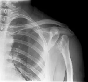
Background imageX Ray Machine Collection: Septic arthritis, X-ray C017 / 7385

Septic arthritis, X-ray C017 / 7385
Septic arthritis. X-ray of the shoulder joint of a 24 year old male that has been damaged due to septic arthritis. Septic arthritis is the inflammation of a joint due to a bacterial infection

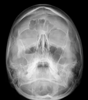
Background imageX Ray Machine Collection: Facial fractures, X-ray C017 / 7638

Facial fractures, X-ray C017 / 7638
Facial fractures. X-ray of the skull of a 25 year old male patient with fractures to the right side (seen here on the left) of his face

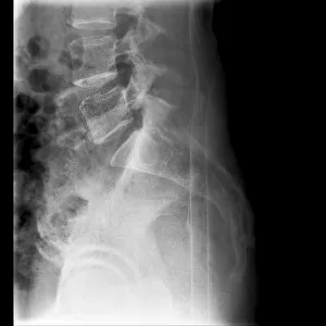
Background imageX Ray Machine Collection: Fractured vertebra, X-ray C017 / 7585

Fractured vertebra, X-ray C017 / 7585
Fractured vertebra. X-ray of the lower spine of a 50 year old male with a fracture to the L1 lumbar vertebra (top centre)

Background imageX Ray Machine Collection: Fractured elbow, X-ray C017 / 7182

Fractured elbow, X-ray C017 / 7182
Fractured elbow. X-ray of the fractured left elbow of a 9 year old boy

Background imageX Ray Machine Collection: Dislocated wrist, X-ray C017 / 7563

Dislocated wrist, X-ray C017 / 7563
Dislocated wrist. X-ray of the wrist of a 16 year old male that has been dislocated

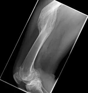
Background imageX Ray Machine Collection: Malunited fracture, X-ray C017 / 7641

Malunited fracture, X-ray C017 / 7641
Malunited fracture. X-ray of the leg of a 64 year old female patient with a malunited fracture of the femur (thigh bone). This occurs where a fracture fails to heal in the correct position

Background imageX Ray Machine Collection: Small bowel obstruction, X-ray C014 / 1819

Small bowel obstruction, X-ray C014 / 1819
Small bowel obstruction. Coloured X-ray of the abdomen of a 77 year old male patient with a small bowel obstruction and gallstones (spheres at top left). The white areas are gas-filled bowel loops

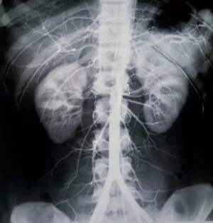
Background imageX Ray Machine Collection: Healthy kidneys, X-ray C014 / 4920

Healthy kidneys, X-ray C014 / 4920
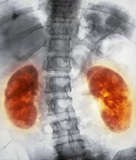
Healthy kidneys. Arteriograph (artery X-ray) of the abdomen showing two healthy kidneys (yellow, left and right). he kidneys filter waste products from the blood

Background imageX Ray Machine Collection: Uterine fibroid, X-ray C014 / 4919

Uterine fibroid, X-ray C014 / 4919
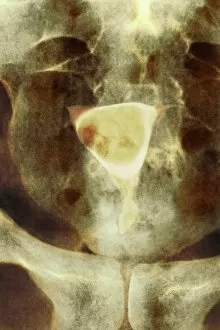
Uterus tumour. Coloured X-ray of the abdomen of a woman with a fibroid (round, centre) in her uterus. A fibroid is a fibrous benign tumour originating from muscular tissue (leimyoma)

Background imageX Ray Machine Collection: Uterine fibroid, X-ray C014 / 4918

Uterine fibroid, X-ray C014 / 4918
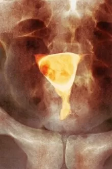
Uterus tumour. Coloured X-ray of the abdomen of a woman with a fibroid (orange, round) in her uterus. A fibroid is a fibrous benign tumour originating from muscular tissue (leimyoma)

Background imageX Ray Machine Collection: Endometriosis, X-ray C014 / 4917

Endometriosis, X-ray C014 / 4917
Endometriosis. Coloured X-ray of the abdomen of woman with endometriosis. Endometriosis is the movement of cells from the lining of the uterus to the abdominal cavity

Background imageX Ray Machine Collection: Endometriosis, X-ray C014 / 4916

Endometriosis, X-ray C014 / 4916
Endometriosis. Coloured X-ray of the abdomen of woman with endometriosis. Endometriosis is the movement of cells from the lining of the uterus to the abdominal cavity

Background imageX Ray Machine Collection: Healthy kidneys, X-ray C014 / 4921

Healthy kidneys, X-ray C014 / 4921
Healthy kidneys. Arteriograph (artery X-ray) of the abdomen showing two healthy kidneys (orange). he kidneys filter waste products from the blood

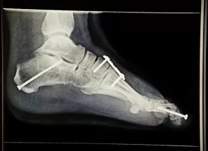
Background imageX Ray Machine Collection: Pinned foot bones after surgery, X-ray

Pinned foot bones after surgery, X-ray
Pinned foot bones after surgery. Lateral (side view) X-ray showing pins (white) inserted into the bones of a patients foot

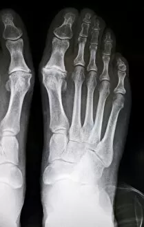
Background imageX Ray Machine Collection: Hallux rigidus before surgery, X-ray

Hallux rigidus before surgery, X-ray
Hallux rigidus before surgery. X-ray showing foot bones before surgery to correct a case of hallux rigidus, a stiff big toe

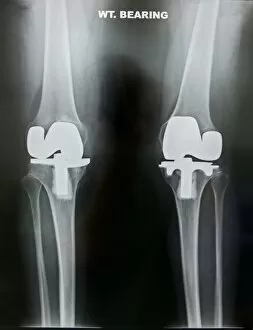
Background imageX Ray Machine Collection: Artificial knee joints in obesity, X-ray

Artificial knee joints in obesity, X-ray
Artificial knee joints in obesity. X-ray of the replacement knee joints of an obese patient. This is a weight-bearing view from the front

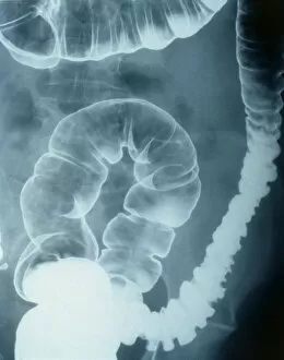
Background imageX Ray Machine Collection: Colonic spasm, X-ray

Colonic spasm, X-ray
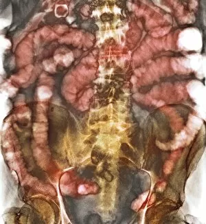
Colonic spasm. Double contrast coloured X-ray of a spasm in the large intestine (colon). The spasm is the narrowed area at left. This could be caused by irritable bowel syndrome

Background imageX Ray Machine Collection: Stomach cancer, barium X-ray

Stomach cancer, barium X-ray
Stomach cancer. Coloured frontal X-ray of the upper gastroduodenal tract of a patient with stomach cancer (grey, upper centre)

Background imageX Ray Machine Collection: Atherosclerosis, X-ray

Atherosclerosis, X-ray
Atherosclerosis. X-ray of a patient with atherosclerosis of the abdominal aorta (white, down centre). The aorta is the bodys main artery, it carries blood from the heart to the rest of the body

Background imageX Ray Machine Collection: Thigh bone head, X-ray

Thigh bone head, X-ray
Thigh bone head. Coloured X-ray of the head of a femur (thigh bone). Seen here is cancellous, or spongy, bone, which is characterised by a honeycomb arrangement comprising a network of trabeculae

Background imageX Ray Machine Collection: Healthy stomach, X-ray

Healthy stomach, X-ray
Healthy stomach, coloured X-ray. The stomach is a curved, sac-like organ that receives food from the oesophagus and passes it to the small intestine

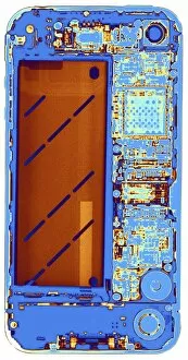
Background imageX Ray Machine Collection: IPhone 4, coloured x-ray C016 / 7214

IPhone 4, coloured x-ray C016 / 7214
Coloured x-ray of an iPhone 4, showing the internal structure. The iPhone 4 mobile phone was introduced on 24 June 2010. It is a smart phone

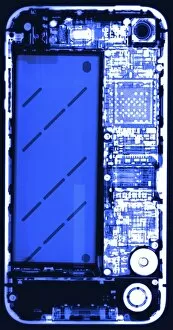
Background imageX Ray Machine Collection: IPhone, X-ray C016 / 7207

IPhone, X-ray C016 / 7207
X-ray of an iPhone 4, showing the internal structure. The iPhone 4 mobile phone was introduced on 24 June 2010. It is a smart phone

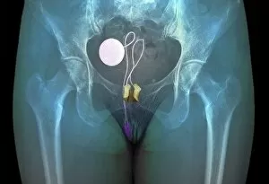
Background imageX Ray Machine Collection: Artificial urinary sphincter, X-ray C016 / 6609

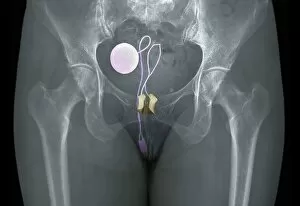
Artificial urinary sphincter, X-ray C016 / 6609
Artificial urinary sphincter. Coloured X-ray of the pelvis of a 55 year old female patient showing a prosthetic urinary sphincter (AUS, beige at centre)

Background imageX Ray Machine Collection: Artificial urinary sphincter, X-ray C016 / 6610

Artificial urinary sphincter, X-ray C016 / 6610
Artificial urinary sphincter. Coloured X-ray of the pelvis of a 55 year old female patient showing a prosthetic urinary sphincter (AUS, beige at centre)

Background imageX Ray Machine Collection: Fractured and dislocated shoulder, X-ray C016 / 6607

Fractured and dislocated shoulder, X-ray C016 / 6607
Fractured and dislocated shoulder. Coloured X-ray of the shoulder of a 67 year old patient who has fallen and fractured the neck of the humerus (upper arm bone) and dislocated the shoulder

Background imageX Ray Machine Collection: Artificial urinary sphincter, X-ray C016 / 6608

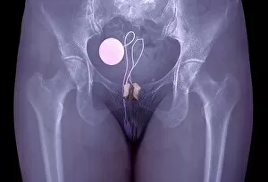
Artificial urinary sphincter, X-ray C016 / 6608
Artificial urinary sphincter. Coloured X-ray of the pelvis of a 55 year old female patient showing a prosthetic urinary sphincter (AUS, beige at centre)

Background imageX Ray Machine Collection: Fractured and dislocated shoulder, X-ray C016 / 6606

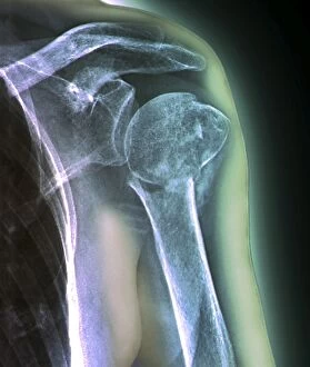
Fractured and dislocated shoulder, X-ray C016 / 6606
Fractured and dislocated shoulder. Coloured X-ray of the shoulder of a 67 year old patient who has fallen and fractured the neck of the humerus (upper arm bone) and dislocated the shoulder

Background imageX Ray Machine Collection: Knee realignment surgery, X-ray C016 / 6604

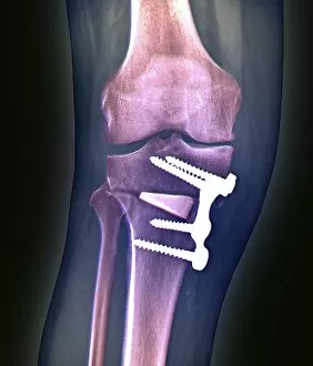
Knee realignment surgery, X-ray C016 / 6604
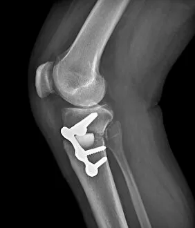
Knee alignment surgery. Coloured X-ray of the knee of a 55 year old overweightpatient that has undergone a wedge osteotomy to correct the alignment of the knee joint

Background imageX Ray Machine Collection: Knee realignment surgery, X-ray C016 / 6601

Knee realignment surgery, X-ray C016 / 6601
Knee alignment surgery. Coloured X-ray of the knee of a 55 year old overweightpatient that has undergone a wedge osteotomy to correct the alignment of the knee joint

Background imageX Ray Machine Collection: Knee realignment surgery, X-ray C016 / 6603

Knee realignment surgery, X-ray C016 / 6603
Knee alignment surgery. Coloured X-ray of the knee of a 55 year old overweightpatient that has undergone a wedge osteotomy to correct the alignment of the knee joint

Background imageX Ray Machine Collection: Knee realignment surgery, X-ray C016 / 6602

Knee realignment surgery, X-ray C016 / 6602
Knee alignment surgery. X-ray of the knee of a 55 year old overweightpatient that has undergone a wedge osteotomy to correct the alignment of the knee joint

Background imageX Ray Machine Collection: Knee realignment surgery, X-ray C016 / 6599

Knee realignment surgery, X-ray C016 / 6599
Knee alignment surgery. Coloured X-ray of the knee of a 55 year old overweightpatient that has undergone a wedge osteotomy to correct the alignment of the knee joint

Background imageX Ray Machine Collection: Prosthetic knee and obesity, X-ray C016 / 6595

Prosthetic knee and obesity, X-ray C016 / 6595
Prosthetic knee and obesity. Coloured X-ray of a prosthetic knee joint (white) in an obese patient. This patient weighs 102 kilograms

Background imageX Ray Machine Collection: Prosthetic knee and obesity, X-ray C016 / 6594

Prosthetic knee and obesity, X-ray C016 / 6594
Prosthetic knee and obesity. Coloured X-ray of a prosthetic knee joint (white) in an obese patient. This patient weighs 102 kilograms

Background imageX Ray Machine Collection: Prosthetic knee and obesity, X-ray C016 / 6593

Prosthetic knee and obesity, X-ray C016 / 6593
Prosthetic knee and obesity. X-ray of a prosthetic knee joint (white) in an obese patient. This patient weighs 102 kilograms

Background imageX Ray Machine Collection: Balloon angioplasty, X-rays C016 / 6589

Balloon angioplasty, X-rays C016 / 6589
Balloon angioplasty. Coloured angiograms (X-rays) of the leg of a 69 year old patient with stenosis (narrowing) of the popliteal artery

Background imageX Ray Machine Collection: Balloon angioplasty, X-ray C016 / 6591

Balloon angioplasty, X-ray C016 / 6591
Balloon angioplasty. Coloured angiogram (X-ray) of the leg of a 69 year old patient undergoing a balloon angioplasty to treat stenosis (narrowing) of the popliteal artery

Background imageX Ray Machine Collection: Balloon angioplasty, X-ray C016 / 6590

Balloon angioplasty, X-ray C016 / 6590
Balloon angioplasty. Angiogram (X-ray) of the leg of a 69 year old patient undergoing a balloon angioplasty to treat stenosis (narrowing) of the popliteal artery

Background imageX Ray Machine Collection: Balloon angioplasty, X-rays C016 / 6587

Balloon angioplasty, X-rays C016 / 6587
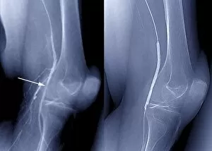
Balloon angioplasty. Coloured angiograms (X-rays) of the leg of a 69 year old patient with stenosis (narrowing, indicated by arrow at left) of the popliteal artery

Background imageX Ray Machine Collection: Pinned leg fracture, X-ray C016 / 6586

Pinned leg fracture, X-ray C016 / 6586
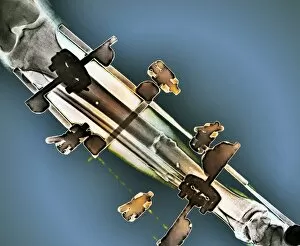
Pinned leg fracture. X-ray of the leg of a 33 year old patient that has been externally fixed after a skiing accident. Both the tibia (shine bone, upper bone) and fibula (calf bone)

Background imageX Ray Machine Collection: Pinned leg fracture, X-ray C016 / 6585

Pinned leg fracture, X-ray C016 / 6585
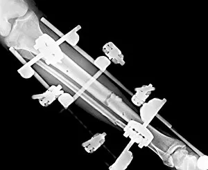
Pinned leg fracture. X-ray of the leg of a 33 year old patient that has been externally fixed after a skiing accident. Both the tibia (shine bone, upper bone) and fibula (calf bone)

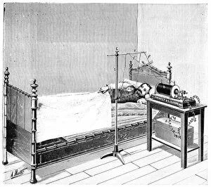
Background imageX Ray Machine Collection: Treatment using X-rays, 1897

Treatment using X-rays, 1897
Treatment using X-rays. 19th-century artwork of a patient in bed being treated by X-rays from the machine at right. At the time (1897)

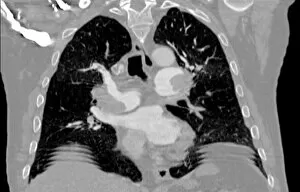
Background imageX Ray Machine Collection: Pulmonary embolism, CT scan

Pulmonary embolism, CT scan
Pulmonary embolism. Computed tomography (CT) scan of a section through a 65 year old male patients chest showing blood clots, or embolisms (white, centre left adn centre right)

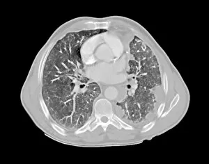
Background imageX Ray Machine Collection: Mesothelioma lung cancer, CT scan

Mesothelioma lung cancer, CT scan
Mesothelioma lung cancer. Computed tomography (CT) scan of the lungs of a 69 year old patient with mesothelioma, a type of lung cancer

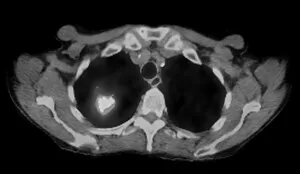
Background imageX Ray Machine Collection: Lung cancer, CT and PET scans

Lung cancer, CT and PET scans
Lung cancer. Combined computed tomography (CT) and positron emission tomography (PET) scans of the lungs of an 84 year old woman with a malignant (cancerous) tumour (bright patch)

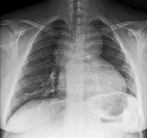
Background imageX Ray Machine Collection: Congenital heart disease, X-ray C017 / 8020

Congenital heart disease, X-ray C017 / 8020
Congenital heart disease. X-ray of the chest of a 19 year old female patient with congenital (present at birth) heart disease


For sale as Licensed Images

Choose your image, Select your licence and Download the media

The incredible versatility of the x-ray machine never ceases to amaze us. From capturing intricate details of brain blood vessels in a 3D angiogram C007 / 1981, to diagnosing a broken wrist bone in X-ray C017 / 7187, this technology has revolutionized medical imaging. But it doesn't stop there. The x-ray machine has even ventured into the animal kingdom, revealing fascinating insights such as a horse's skull or the delicate skeleton of a seahorse. It seems that no creature is too small or too large for this remarkable tool. In addition to its medical applications, the x-ray machine also showcases its playful side. Who would have thought that skeletons could engage in sports? A rugby-playing skeleton and even one enjoying a refreshing drink are just some how this technology can bring humor and creativity to our lives. Not limited to humans and animals, the x-ray machine extends its reach into other fields as well. Panoramic dental X-rays help dentists assess oral health with precision while an individual holding a camera reveals how this invention itself can be captured on film. Furthermore, we witness how the x-ray machine aids in surgical procedures like total hip replacements by providing surgeons with detailed images for precise placement and alignment. Last but not least, let's not forget about exploring what lies beneath our own skin - normal skulls are examined through X-rays allowing us to marvel at the intricacies hidden within each one of us. From healthcare advancements to artistic expressions and scientific discoveries, these glimpses into various aspects of life demonstrate why the x-ray machine remains an invaluable tool that continues to shape our understanding of both human anatomy and beyond.