X Ray Machine Collection (#7)

The incredible versatility of the x-ray machine never ceases to amaze us

Background imageX Ray Machine Collection: Balloon angioplasty, X-ray C016 / 6590

Balloon angioplasty, X-ray C016 / 6590
Balloon angioplasty. Angiogram (X-ray) of the leg of a 69 year old patient undergoing a balloon angioplasty to treat stenosis (narrowing) of the popliteal artery

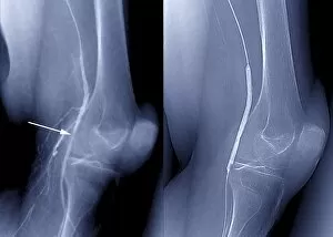
Background imageX Ray Machine Collection: Balloon angioplasty, X-rays C016 / 6587

Balloon angioplasty, X-rays C016 / 6587
Balloon angioplasty. Coloured angiograms (X-rays) of the leg of a 69 year old patient with stenosis (narrowing, indicated by arrow at left) of the popliteal artery

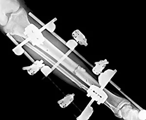
Background imageX Ray Machine Collection: Pinned leg fracture, X-ray C016 / 6586

Pinned leg fracture, X-ray C016 / 6586
Pinned leg fracture. X-ray of the leg of a 33 year old patient that has been externally fixed after a skiing accident. Both the tibia (shine bone, upper bone) and fibula (calf bone)

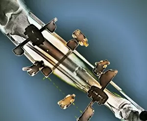
Background imageX Ray Machine Collection: Pinned leg fracture, X-ray C016 / 6585

Pinned leg fracture, X-ray C016 / 6585
Pinned leg fracture. X-ray of the leg of a 33 year old patient that has been externally fixed after a skiing accident. Both the tibia (shine bone, upper bone) and fibula (calf bone)

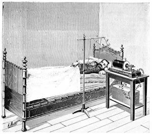
Background imageX Ray Machine Collection: Treatment using X-rays, 1897

Treatment using X-rays, 1897
Treatment using X-rays. 19th-century artwork of a patient in bed being treated by X-rays from the machine at right. At the time (1897)

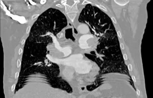
Background imageX Ray Machine Collection: Pulmonary embolism, CT scan

Pulmonary embolism, CT scan
Pulmonary embolism. Computed tomography (CT) scan of a section through a 65 year old male patients chest showing blood clots, or embolisms (white, centre left adn centre right)

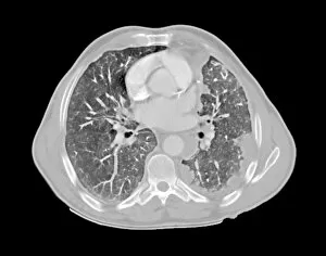
Background imageX Ray Machine Collection: Mesothelioma lung cancer, CT scan

Mesothelioma lung cancer, CT scan
Mesothelioma lung cancer. Computed tomography (CT) scan of the lungs of a 69 year old patient with mesothelioma, a type of lung cancer

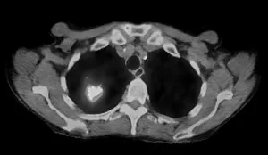
Background imageX Ray Machine Collection: Lung cancer, CT and PET scans

Lung cancer, CT and PET scans
Lung cancer. Combined computed tomography (CT) and positron emission tomography (PET) scans of the lungs of an 84 year old woman with a malignant (cancerous) tumour (bright patch)

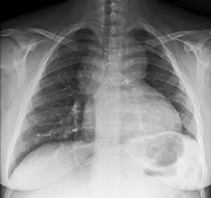
Background imageX Ray Machine Collection: Congenital heart disease, X-ray C017 / 8020

Congenital heart disease, X-ray C017 / 8020
Congenital heart disease. X-ray of the chest of a 19 year old female patient with congenital (present at birth) heart disease

Background imageX Ray Machine Collection: Salter Harris fracture, X-ray C017 / 8025

Salter Harris fracture, X-ray C017 / 8025
Salter Harris fracture. X-ray of the ankle of a child patient with a displaced Salter Harris fracture of the ankle. Salter Harris fractures occur at the epiphyseal, or growth

Background imageX Ray Machine Collection: Metal in eye, X-ray C017 / 8019

Metal in eye, X-ray C017 / 8019
Metal in eye. X-ray of the face of a 64 year old male patient showing metal in the orbit of the eye

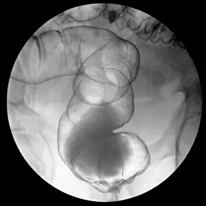
Background imageX Ray Machine Collection: Diverticulitis C017 / 8015

Diverticulitis C017 / 8015
Diverticulitis. X-ray of the colon of a patient with diverticulitis. A diverticulum is a small pouch-like structure that protrudes out of weak spots in the intestinal wall

Background imageX Ray Machine Collection: Diverticulitis C017 / 8014

Diverticulitis C017 / 8014
Diverticulitis. X-ray of the colon of a patient with diverticulitis. A diverticulum is a small pouch-like structure that protrudes out of weak spots in the intestinal wall

Background imageX Ray Machine Collection: Arthritic knees, X-ray C017 / 8013

Arthritic knees, X-ray C017 / 8013
Arthritic knees. X-ray of the knees of a patient with osteoarthritis. Osteoarthritis is a joint disease caused by mechanical wear and tear

Background imageX Ray Machine Collection: Ankle fracture with bone cyst, X-ray C017 / 8016

Ankle fracture with bone cyst, X-ray C017 / 8016
Ankle fracture. X-ray of the fractured ankle of a patient. The bone had been weakened by a cyst, a fluid-filled cavity

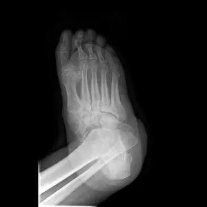
Background imageX Ray Machine Collection: Crushed right foot, X-ray C017 / 8012

Crushed right foot, X-ray C017 / 8012
Crushed right foot. X-ray of the right foot of an 18 year old male patient who has been in a motorcycle accident

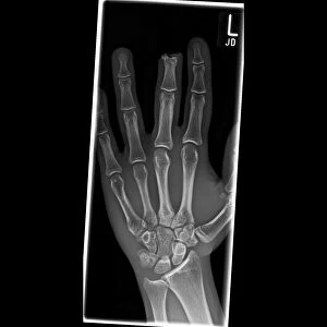
Background imageX Ray Machine Collection: Amputated finger, X-ray C017 / 8010

Amputated finger, X-ray C017 / 8010
Amputated finger. X-ray of the hand of a 17 year old male patient who has had the tip of his middle finger amputated

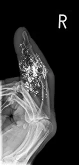
Background imageX Ray Machine Collection: Foreign body in finger, X-ray C017 / 8009

Foreign body in finger, X-ray C017 / 8009
Foreign body in finger. X-ray of the finger of a 60 year old male patient that contains a foreign body that is causing swelling of the soft tissue

Background imageX Ray Machine Collection: Broken wrist, X-ray C017 / 8008

Broken wrist, X-ray C017 / 8008
Broken wrist. X-ray of the arm of an 84 year old female patient with a fractured wrist

Background imageX Ray Machine Collection: Extra fingers, X-ray C017 / 8007

Extra fingers, X-ray C017 / 8007
Extra fingers. X-ray of the hand of a one year old male patient with an extra finger, a congenital (present at birth) abnormality known as polydactyly

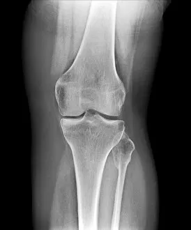
Background imageX Ray Machine Collection: Removed kneecap, X-ray C017 / 7981

Removed kneecap, X-ray C017 / 7981
Removed kneecap. X-ray of the knee of a 46 year old female that has had her kneecap (patella) surgically removed in a procedure known as a patellectomy

Background imageX Ray Machine Collection: Pinned jaw fracture, X-ray C017 / 7982

Pinned jaw fracture, X-ray C017 / 7982
Pinned jaw fracture. X-ray of the head of a 22 year old male patient who has had a fractured mandible (lower jaw bone) pinned (lower left)

Background imageX Ray Machine Collection: Duplex kidney collecting system, X-ray C017 / 7973

Duplex kidney collecting system, X-ray C017 / 7973
Duplex kidney collecting system. X-ray of the abdomen of a 78 year old male patient who has bilateral duplex collecting systems of the kidneys

Background imageX Ray Machine Collection: Nail in foot, X-ray C017 / 7980

Nail in foot, X-ray C017 / 7980
Nail in foot. X-ray of the foot of a 7 year old female patient who had stood on a nail

Background imageX Ray Machine Collection: Broken elbow, X-ray C017 / 7979

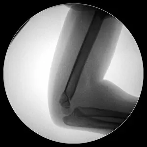
Broken elbow, X-ray C017 / 7979
Broken elbow. X-ray of the elbow of a 4 year old female with a displaced supracondylar fracture. The fracture is at the base of the humerus, the upper arm bone (upper frame)

Background imageX Ray Machine Collection: Pinned broken ankle, X-ray C017 / 7978

Pinned broken ankle, X-ray C017 / 7978
Pinned broken ankle. X-ray of the ankle of a 32 year old male that has been pinned after a parachuting accident. The ankle was dislocated between the tibia (shin bone)

Background imageX Ray Machine Collection: Fractured and dislocated ankle, X-ray C017 / 7976

Fractured and dislocated ankle, X-ray C017 / 7976
Fractured and dislocated ankle. X-ray of the ankle of a 32 year old male who has been in a parachuting accident. The ankle is dislocated between the tibia (shin bone)

Background imageX Ray Machine Collection: Infected malunited fracture, X-ray C017 / 7972

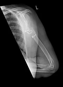
Infected malunited fracture, X-ray C017 / 7972
Infected malunited fracture. X-ray of the arm of a 65 year old female patient who has a bone infection after the nonunion of a fracture of the humerus (upper arm bone)

Background imageX Ray Machine Collection: Pneumothorax, X-ray C017 / 7966

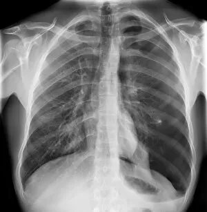
Pneumothorax, X-ray C017 / 7966
Pneumothorax. X-ray of the chest of a 22 year old male patient with a pneumothorax, or collapsed lung. The left lung (right on X-ray) has collapsed due to a build-up of air (solid grey)

Background imageX Ray Machine Collection: Knuckle replacement, X-ray C017 / 7971

Knuckle replacement, X-ray C017 / 7971
Knuckle replacement. X-ray of the hand of a 41 year old male patient who has had two of the joints replaced between the hand bones (metacarpals) and the first finger bones (proximal phalanges)

Background imageX Ray Machine Collection: Broken elbow, X-ray C017 / 7968

Broken elbow, X-ray C017 / 7968
Broken elbow. X-ray of the elbow of a 5 year old female with a displaced supracondylar fracture. The fracture is at the base of the humerus, the upper arm bone (upper frame)

Background imageX Ray Machine Collection: Knuckle replacement, X-ray C017 / 7970

Knuckle replacement, X-ray C017 / 7970
Knuckle replacement. X-ray of the hand of a 41 year old male patient who has had two of the joints replaced between the hand bones (metacarpals) and the first finger bones (proximal phalanges)

Background imageX Ray Machine Collection: Pinned fractured elbow, X-ray C017 / 7969

Pinned fractured elbow, X-ray C017 / 7969
Pinned fractured elbow. X-ray of the elbow of a 5 year old female that has been internally fixed after a displaced supracondylar fracture

Background imageX Ray Machine Collection: Reversed heart position, X-ray C017 / 7965

Reversed heart position, X-ray C017 / 7965
Reversed heart position. X-ray of the chest of a patient with dextrocardia, the congenital (present at birth) placement of the heart on the right side of the body

Background imageX Ray Machine Collection: Arthritic hands, X-ray C017 / 7967

Arthritic hands, X-ray C017 / 7967
Arthritic hands. X-ray of the hands of a 49 year old female patient with arthritis of the finger joints. Arthritis is a degenerative disease that causes the loss of the protective cartilage between

Background imageX Ray Machine Collection: Lung cancer, X-ray C017 / 7964

Lung cancer, X-ray C017 / 7964
Lung cancer. X-ray if the chest of a patient with malignant (cancerous) tumours in middle of the left lung (seen at right). The main cause of lung cancer is cigarette smoke

Background imageX Ray Machine Collection: Knife in hand, X-ray C017 / 7963

Knife in hand, X-ray C017 / 7963
Knife in hand. X-ray of a knife through the left hand of a patient
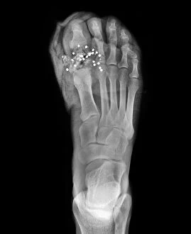
Background imageX Ray Machine Collection: Firearm injury, X-ray C017 / 7873

Firearm injury, X-ray C017 / 7873
Firearm injury. X-ray of the foot of a patient who has been shot with a shotgun. Multiple pellets (white dots) can be seen in the big toe
Background imageX Ray Machine Collection: Gallstones, X-ray C017 / 7860

Gallstones, X-ray C017 / 7860
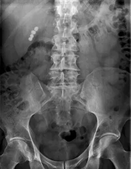
Gallstones. X-ray of the abdomen of a patient showing four gallstones (white balls at upper left). Gallstones (choleliths)

Background imageX Ray Machine Collection: Bone infection, X-ray C017 / 7861

Bone infection, X-ray C017 / 7861
Bone infection. X-ray of the foot of a diabetic patient osteomyelitis, a bacterial infection of the bone. Tissue damage to the foot caused by diabetes has lead to the infection

Background imageX Ray Machine Collection: Ankle fracture with bone cyst, X-ray C017 / 7862

Ankle fracture with bone cyst, X-ray C017 / 7862
Ankle fracture. X-ray of the fractured ankle of a patient. The bone had been weakened by a cyst, a fluid-filled cavity

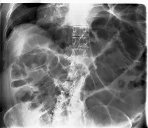
Background imageX Ray Machine Collection: Large bowel obstruction, X-ray C017 / 7855

Large bowel obstruction, X-ray C017 / 7855
Large bowel obstruction. X-ray of the abdomen of a patient with an obstructed large intestine. The obstruction may be due to cancer, twisting of the intestine, an injury or an infection

Background imageX Ray Machine Collection: Large bowel obstruction, X-ray C017 / 7854

Large bowel obstruction, X-ray C017 / 7854
Large bowel obstruction. X-ray of the abdomen of a patient with an obstructed large intestine. The obstruction may be due to cancer, twisting of the intestine, an injury or an infection

Background imageX Ray Machine Collection: Kidney stones, X-ray C017 / 7856

Kidney stones, X-ray C017 / 7856
Kidney stones. X-ray of the kidney of a patient with kidney stones (dark areas) in the renal pelvis (upper centre) of the kidney

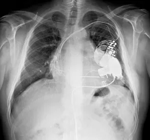
Background imageX Ray Machine Collection: Pacemaker and heart pump, X-ray C017 / 7833

Pacemaker and heart pump, X-ray C017 / 7833
Pacemaker and heart pump. X-ray of the chest of a patient who is waiting for a heart and lung transplant. They have a pacemaker and a left ventricular assist device (LVAD) fitted

Background imageX Ray Machine Collection: Diverticulitis C017 / 7835

Diverticulitis C017 / 7835
Diverticulitis. X-ray of the colon of a patient with diverticulitis. A diverticulum is a small pouch-like structure that protrudes out of weak spots in the intestinal wall

Background imageX Ray Machine Collection: Enlarged heart, X-ray C017 / 7830

Enlarged heart, X-ray C017 / 7830
Enlarged heart. X-ray of the chest of a female patient with an enlarged heart. Cardiac diseases can make the heart need to pump harder

Background imageX Ray Machine Collection: Enlarged heart, X-ray C017 / 7827

Enlarged heart, X-ray C017 / 7827
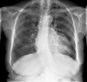
Enlarged heart. X-ray of the chest of a patient with an enlarged heart and a pacemaker (top right). Cardiac diseases can make the heart need to pump harder


For sale as Licensed Images

Choose your image, Select your licence and Download the media

The incredible versatility of the x-ray machine never ceases to amaze us. From capturing intricate details of brain blood vessels in a 3D angiogram C007 / 1981, to diagnosing a broken wrist bone in X-ray C017 / 7187, this technology has revolutionized medical imaging. But it doesn't stop there. The x-ray machine has even ventured into the animal kingdom, revealing fascinating insights such as a horse's skull or the delicate skeleton of a seahorse. It seems that no creature is too small or too large for this remarkable tool. In addition to its medical applications, the x-ray machine also showcases its playful side. Who would have thought that skeletons could engage in sports? A rugby-playing skeleton and even one enjoying a refreshing drink are just some how this technology can bring humor and creativity to our lives. Not limited to humans and animals, the x-ray machine extends its reach into other fields as well. Panoramic dental X-rays help dentists assess oral health with precision while an individual holding a camera reveals how this invention itself can be captured on film. Furthermore, we witness how the x-ray machine aids in surgical procedures like total hip replacements by providing surgeons with detailed images for precise placement and alignment. Last but not least, let's not forget about exploring what lies beneath our own skin - normal skulls are examined through X-rays allowing us to marvel at the intricacies hidden within each one of us. From healthcare advancements to artistic expressions and scientific discoveries, these glimpses into various aspects of life demonstrate why the x-ray machine remains an invaluable tool that continues to shape our understanding of both human anatomy and beyond.