
Zoonosis Collection
Zoonosis: Unveiling the Microscopic World of Infectious Threats In a world teeming with microscopic organisms


For sale as Licensed Images
Choose your image, Select your licence and Download the media
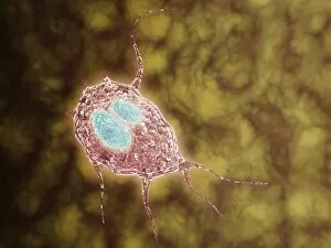
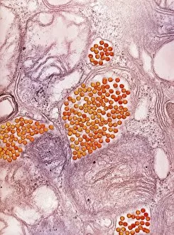
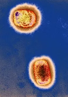
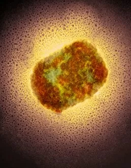
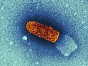
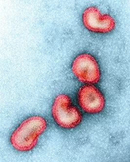
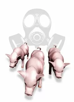
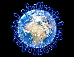
Zoonosis: Unveiling the Microscopic World of Infectious Threats In a world teeming with microscopic organisms, zoonosis emerges as a formidable threat to both animal and human health. With its ability to jump across species barriers, this phenomenon has captured the attention of scientists worldwide. Delving into the microscopic view of various zoonotic agents, we uncover their potential for causing widespread diseases. Giardiasis, caused by Giardia parasites, reveals its intricate structure under close examination. This common waterborne illness affects both humans and animals alike, highlighting the ease with which zoonotic infections can spread through contaminated sources. Eastern equine encephalitis virus (EEEV) takes center stage in our exploration. Its presence is magnified through transmission electron microscopy (TEM), showcasing its complex composition that poses a severe threat to horses and occasionally humans. Artwork depicting Lassa virus particles captures our imagination as we witness their elegant yet deadly form. Responsible for Lassa fever outbreaks primarily in West Africa, these viruses exemplify how zoonoses can cause devastating epidemics if left unchecked. Rift Valley fever virus particles also make an appearance within our visual journey into the realm of zoonosis. Their distinct shape serves as a reminder that livestock and mosquitoes act as key players in spreading this viral disease throughout Africa and parts of Asia. Monkeypox virus particles reveal themselves under TEM's watchful eye – tiny entities capable of triggering outbreaks reminiscent of smallpox but less severe in nature. These images emphasize how even seemingly innocuous creatures like rodents can harbor dangerous pathogens that threaten human populations. As we unravel the mysteries hidden within these microscopic views, it becomes evident that zoonoses transcend borders and species boundaries effortlessly. The urgency to understand their mechanisms intensifies as humanity strives to prevent future pandemics from emerging due to such infectious agents lurking among us.