
Zoonotic Collection
Zoonotic diseases pose a significant threat to both humans and animals, highlighting the importance of understanding and preventing their transmission


For sale as Licensed Images
Choose your image, Select your licence and Download the media
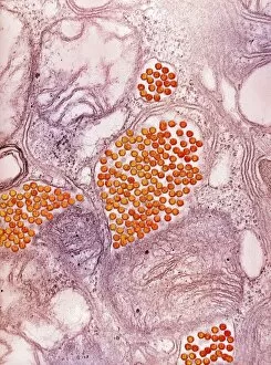
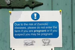
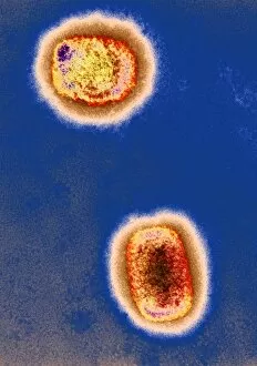
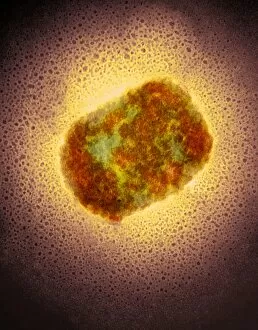
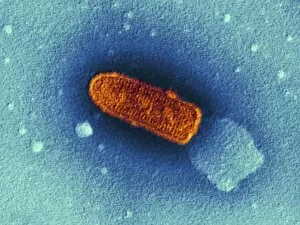
Zoonotic diseases pose a significant threat to both humans and animals, highlighting the importance of understanding and preventing their transmission. One such example is the Eastern equine encephalitis virus, which can cause severe brain inflammation in infected individuals. Through Transmission Electron Microscopy (TEM), scientists have been able to visualize this dangerous pathogen, raising awareness about its structure and potential impact. Due to the risk diseases like Eastern equine encephalitis, it is crucial to take necessary precautions when visiting farms or areas where these viruses may be present. Pregnant women or those suspecting pregnancy should refrain from entering such environments as they are particularly vulnerable. Another zoonotic disease that demands attention is Lassa fever caused by the Lassa virus. Artwork depicting this viral particle showcases its intricate structure, serving as a reminder of the need for vigilance against this deadly pathogen. Similarly, Rift Valley fever virus particles also pose a significant health concern and require proactive measures for prevention. The presence diseases like Lassa fever necessitates strict adherence to safety protocols on farms or in regions where these viruses are prevalent. The artwork displaying various stages of Lassa virus particles serves as a visual representation of the danger they possess. Moreover, Monkeypox virus particles captured through TEM further emphasize the risks associated with zoonoses. These microscopic images highlight how easily these pathogens can spread between animals and humans if proper precautions are not taken. Understanding zoonotic diseases like Eastern equine encephalitis, Lassa fever, and Monkeypox is essential for safeguarding public health. By being aware of their existence and taking appropriate preventive measures such as avoiding high-risk environments during pregnancy or suspected pregnancies, we can minimize the chances of contracting these potentially life-threatening infections.