Metal Print > Popular Themes > Human Body
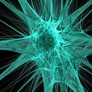
Metal Print : Nerve and glial cells, light micrograph


![]()
Metal Prints from Science Photo Library
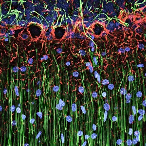
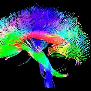
Nerve and glial cells, light micrograph
Nerve and glial cells, fluorescence light micrograph. These are neural stem cells that have differentiated into neurons (nerve cells, blue) and glial cells (support cells, red). The branching processes from the neurons are called dendrites. Fluorescent markers have been used to highlight proteins. The proteins stained here are beta III-tubulin (blue), a cytoskeleton element found in neurons, and GFAP (glial fibrillary acidic protein, red), forming the cytoskeleton of the glial cells. This sample is from rat tissue
Science Photo Library features Science and Medical images including photos and illustrations
Media ID 10948201
© DANIEL SCHROEN, CELL APPLICATIONS INC/SCIENCE PHOTO LIBRARY
Animal Body Astrocyte Astrocytes Cell Biology Cellular Cytoskeletal Cytoskeleton Fluorescence Fluorescence Micrograph Fluorescing Gfap Glial Cell Glial Fibrillary Acidic Protein Nerve Nerve Cell Neuron Neurone Neurones Neurons Nobody Proteins Stains Tubulin Brain Cells Light Micrograph Light Microscope Nervous System Neurological Neurology Protein
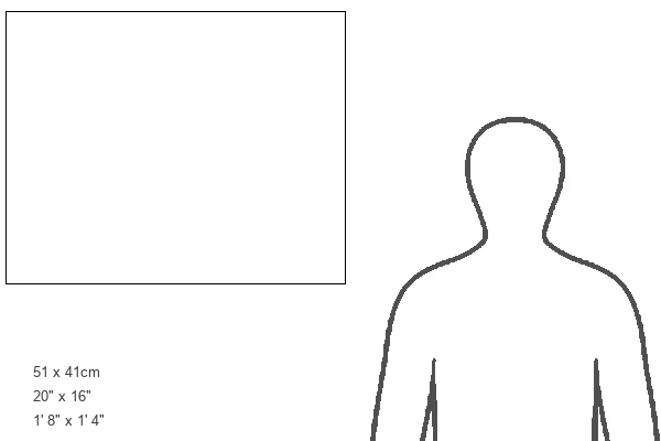
16"x20" (51x41cm) Metal Print
Bring the intricacies of the natural world into your home or office with our Media Storehouse Metal Prints. This stunning image showcases the beauty of nerve and glial cells, captured in a fluorescence light micrograph by Daniel Schroen of Cell Applications Inc/Science Photo Library. Witness the mesmerizing dance of differentiated neural stem cells, as they transform into neurons (blue) and glial cells (red), providing essential support to the nervous system. Our high-quality metal prints offer vibrant colors and exceptional detail, making this a must-have piece for any science enthusiast or modern decor. Experience the wonder of science in a whole new way with Media Storehouse.
Made with durable metal and luxurious printing techniques, our metal photo prints go beyond traditional canvases, adding a cool, modern touch to your space. Wall mount on back. Eco-friendly 100% post-consumer recycled ChromaLuxe aluminum surface. The thickness of the print is 0.045". Featuring a Scratch-resistant surface and Rounded corners. Backing hangers are attached to the back of the print and float the print 1/2-inch off the wall when hung, the choice of hanger may vary depending on size and International orders will come with Float Mount hangers only. Finished with a brilliant white high gloss surface for unsurpassed detail and vibrance. Printed using Dye-Sublimation and for best care we recommend a non-ammonia glass cleaner, water, or isopropyl (rubbing) alcohol to prevent harming the print surface. We recommend using a clean, lint-free cloth to wipe off the print. The ultra-hard surface is scratch-resistant, waterproof and weatherproof. Avoid direct sunlight exposure.
Made with durable metal and luxurious printing techniques, metal prints bring images to life and add a modern touch to any space
Estimated Image Size (if not cropped) is 50.8cm x 40.6cm (20" x 16")
Estimated Product Size is 51.4cm x 41.2cm (20.2" x 16.2")
These are individually made so all sizes are approximate
Artwork printed orientated as per the preview above, with landscape (horizontal) orientation to match the source image.
EDITORS COMMENTS
This print showcases the intricate world of nerve and glial cells, captured through a fluorescence light micrograph. The neural stem cells in this image have undergone differentiation, transforming into neurons (nerve cells) depicted in blue, as well as glial cells (support cells) shown in red. The dendrites extending from the neurons are responsible for transmitting electrical signals throughout the nervous system. To enhance visibility and highlight specific proteins, fluorescent markers were employed during sample preparation. In this particular image, beta III-tubulin is stained blue to emphasize its presence within neuronal cytoskeletons. Meanwhile, GFAP (glial fibrillary acidic protein), which forms the structural framework of glial cells' cytoskeletons, appears vibrant red. It's important to note that this sample originates from rat tissue and offers valuable insights into cellular biology and neurology research. By studying these fundamental building blocks of our nervous system – astrocytes, neurones, differentiated stem cells – scientists gain a deeper understanding of how our brains function. The photographer behind this remarkable image is Daniel Schroen from Cell Applications Inc/Science Photo Library. This visually striking photograph not only captures the beauty found within biological structures but also serves as a testament to human curiosity and scientific exploration in unraveling the mysteries of life itself.
MADE IN THE USA
Safe Shipping with 30 Day Money Back Guarantee
FREE PERSONALISATION*
We are proud to offer a range of customisation features including Personalised Captions, Color Filters and Picture Zoom Tools
SECURE PAYMENTS
We happily accept a wide range of payment options so you can pay for the things you need in the way that is most convenient for you
* Options may vary by product and licensing agreement. Zoomed Pictures can be adjusted in the Cart.