Metal Print > Science > SEM
Metal Print : SEM of section through human skin

![]()
Metal Prints from Science Photo Library
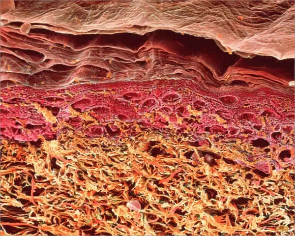
SEM of section through human skin
Skin. Coloured scanning electron micrograph of a section through the human skin. The uppermost layers of the skin (pink and red) make up the epidermis. The surface of the epidermis (at top, pink) is the cornified layer composed of flattened, fused keratin-rich cell remnants. The dead cells from this layer are continuously being shed and replaced from cells from the living epidermal layers below (red). These layers contain keratinocytes, specialized cells that synthesize keratin. The lowest layer shown here is the dermis (yellow). The dermis is a thick layer of fibrous connective tissue which supports and nourishes the epidermis. Magnification: unknown
Science Photo Library features Science and Medical images including photos and illustrations
Media ID 6423286
© STEVE GSCHMEISSNER/SCIENCE PHOTO LIBRARY
Cornified Layer Dermis Epidermis Layers Magnified Image Microscopic Photos Skin Stratum Corneum Subjects Surface
16"x20" (51x41cm) Metal Print
Discover the intricacy of the human body with our Media Storehouse Metal Prints featuring this captivating Scanning Electron Micrograph from Science Photo Library. This mesmerizing image showcases a section through the human skin, revealing the intricate structure of the epidermis in vibrant colors. Our high-quality Metal Prints bring science to life, transforming your space into a gallery of biological wonders. Experience the detail and depth of this SEM image as if you were looking through a microscope, with the added benefit of a sleek, modern design. Perfect for scientists, educators, or anyone with a curiosity for the world around us. Order now and bring the beauty of science into your home or office.
Made with durable metal and luxurious printing techniques, our metal photo prints go beyond traditional canvases, adding a cool, modern touch to your space. Wall mount on back. Eco-friendly 100% post-consumer recycled ChromaLuxe aluminum surface. The thickness of the print is 0.045". Featuring a Scratch-resistant surface and Rounded corners. Backing hangers are attached to the back of the print and float the print 1/2-inch off the wall when hung, the choice of hanger may vary depending on size and International orders will come with Float Mount hangers only. Finished with a brilliant white high gloss surface for unsurpassed detail and vibrance. Printed using Dye-Sublimation and for best care we recommend a non-ammonia glass cleaner, water, or isopropyl (rubbing) alcohol to prevent harming the print surface. We recommend using a clean, lint-free cloth to wipe off the print. The ultra-hard surface is scratch-resistant, waterproof and weatherproof. Avoid direct sunlight exposure.
Made with durable metal and luxurious printing techniques, metal prints bring images to life and add a modern touch to any space
Estimated Image Size (if not cropped) is 50.8cm x 40.6cm (20" x 16")
Estimated Product Size is 51.4cm x 41.2cm (20.2" x 16.2")
These are individually made so all sizes are approximate
Artwork printed orientated as per the preview above, with landscape (horizontal) orientation to match the source image.
FEATURES IN THESE COLLECTIONS
EDITORS COMMENTS
This print offers a mesmerizing glimpse into the intricate structure of human skin. Through the lens of a scanning electron microscope, we are presented with an extraordinary view of the various layers that compose this vital organ. The vibrant colors in this image highlight the uppermost layers known as the epidermis. The pink and red hues represent different regions within this layer. At the top, we can observe the cornified layer, which forms a protective barrier on our skin's surface. Composed of flattened and fused keratin-rich cell remnants, it shields us from external elements. Interestingly, this cornified layer is continuously shedding dead cells while simultaneously replenishing itself with new ones from below. These living epidermal layers (depicted in red) contain specialized cells called keratinocytes that synthesize keratin - a protein crucial for maintaining skin strength and elasticity. Beneath these remarkable epidermal layers lies the dermis, showcased here in yellow tones. This thick layer consists of fibrous connective tissue responsible for supporting and nourishing the overlying epidermis. With its unknown magnification level, this photograph invites us to marvel at nature's intricacy on a microscopic scale. It serves as a reminder of how our bodies are composed of awe-inspiring structures working harmoniously to protect and sustain us.
MADE IN THE USA
Safe Shipping with 30 Day Money Back Guarantee
FREE PERSONALISATION*
We are proud to offer a range of customisation features including Personalised Captions, Color Filters and Picture Zoom Tools
SECURE PAYMENTS
We happily accept a wide range of payment options so you can pay for the things you need in the way that is most convenient for you
* Options may vary by product and licensing agreement. Zoomed Pictures can be adjusted in the Cart.