Mouse Mat > Popular Themes > Human Body
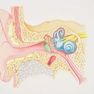
Mouse Mat : Diagram of inner ear showing auditory canal, eardrum, semicircular canals, cochlea, cochlea nerve, eustachian tube

![]()
Home Decor from Fine Art Storehouse
Diagram of inner ear showing auditory canal, eardrum, semicircular canals, cochlea, cochlea nerve, eustachian tube
Unleash your creativity and transform your space into a visual masterpiece!
Dorling Kindersley
Media ID 13560055
© This content is subject to copyright
Anatomy Biology Biomedical Illustration Canals Cochlea Cross Section Diagram Ear Canal Ear Drum Ears Eustachian Tube Function Health Healthcare And Medicine Hearing Human Beings Human Body Human Ear Human Nervous System Humans Listening Model Sensory Perception Sound Square Image Structure Tubes Auditory Eardrum Human Body Part Inner Inner Ear Nerves Perception
Mouse Pad
Standard Size Mouse Pad 7.75" x 9..25". High density Neoprene w linen surface. Easy to clean, stain resistant finish. Rounded corners.
Archive quality photographic print in a durable wipe clean mouse mat with non slip backing. Works with all computer mice
Estimated Product Size is 23.7cm x 20.2cm (9.3" x 8")
These are individually made so all sizes are approximate
Artwork printed orientated as per the preview above, with landscape (horizontal) orientation to match the source image.
FEATURES IN THESE COLLECTIONS
> Fine Art Storehouse
> Photo Libraries
> Dorling Kindersley Prints
EDITORS COMMENTS
This print showcases a detailed diagram of the inner ear, providing an insightful glimpse into the intricate workings of our auditory system. Against a crisp white background, this square image features a cross-section view that highlights various essential components. From left to right, we can observe the auditory canal leading to the eardrum, followed by the semicircular canals and cochlea nerve. The illustration beautifully captures both the structure and function of these vital elements. The cochlea, resembling a delicate seashell, stands out with its spiral shape and is responsible for converting sound vibrations into electrical signals that our brain can interpret as meaningful sounds. Meanwhile, the eustachian tube connects to the back of our throat and helps regulate pressure within the middle ear. With its vibrant colors and precise detailing, this biomedical illustration serves as an invaluable resource for anyone interested in understanding how humans perceive sound. Whether you are studying anatomy or simply curious about your own sensory perception capabilities, this print offers a visually engaging way to explore one of our most fascinating senses – hearing. Created by Dorling Kindersley and available through Fine Art Storehouse, this stunning artwork adds both educational value and aesthetic appeal to any space it graces.
MADE IN THE USA
Safe Shipping with 30 Day Money Back Guarantee
FREE PERSONALISATION*
We are proud to offer a range of customisation features including Personalised Captions, Color Filters and Picture Zoom Tools
SECURE PAYMENTS
We happily accept a wide range of payment options so you can pay for the things you need in the way that is most convenient for you
* Options may vary by product and licensing agreement. Zoomed Pictures can be adjusted in the Cart.