Mouse Mat : Smooth endoplasmic reticulum, SEM

![]()
Home Decor from Science Photo Library
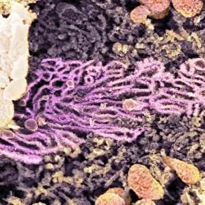
Smooth endoplasmic reticulum, SEM
Endoplasmic reticulum. Coloured scanning electron micrograph (SEM) of smooth endoplasmic reticulum (SER) (orange, centre left) in the cytoplasm of a kidney cell. SER is a membrane-bound cell organelle that is the site of lipid synthesis and the production of membrane-bound proteins
Science Photo Library features Science and Medical images including photos and illustrations
Media ID 6303491
© DR DAVID FURNESS, KEELE UNIVERSITY/SCIENCE PHOTO LIBRARY
Cell Biology Cross Section Cytology Cytoplasm Eukaryotic Kidney Organelle Renal Smooth Endoplasmic Reticulum False Coloured Section Sectioned
Mouse Pad
Standard Size Mouse Pad 7.75" x 9..25". High density Neoprene w linen surface. Easy to clean, stain resistant finish. Rounded corners.
Archive quality photographic print in a durable wipe clean mouse mat with non slip backing. Works with all computer mice
Estimated Image Size (if not cropped) is 23.7cm x 17.4cm (9.3" x 6.9")
Estimated Product Size is 23.7cm x 20.2cm (9.3" x 8")
These are individually made so all sizes are approximate
Artwork printed orientated as per the preview above, with landscape (horizontal) orientation to match the source image.
EDITORS COMMENTS
This print from Science Photo Library showcases the intricate beauty of a smooth endoplasmic reticulum (SER) within a kidney cell. The image, captured using a scanning electron microscope (SEM), reveals the vibrant orange-colored SER located at the center left of the cytoplasm. The smooth endoplasmic reticulum is an essential membrane-bound organelle responsible for various cellular functions. It serves as both the site for lipid synthesis and production of membrane-bound proteins. Its presence in this kidney cell highlights its crucial role in maintaining cellular homeostasis and functionality. The false-colored SEM image provides us with a unique glimpse into the microscopic world of biology. By sectioning and capturing this cross-section view, we can appreciate the complexity and organization present within eukaryotic cells. As we delve deeper into cytology, it becomes evident that every component plays a vital part in sustaining life. This particular photograph emphasizes not only the significance of SER but also underscores how advancements in technology enable us to explore these minute structures with astonishing detail. Science Photo Library continues to provide invaluable resources like this print, enabling researchers, educators, and enthusiasts alike to marvel at nature's wonders on both macroscopic and microscopic scales.
MADE IN THE USA
Safe Shipping with 30 Day Money Back Guarantee
FREE PERSONALISATION*
We are proud to offer a range of customisation features including Personalised Captions, Color Filters and Picture Zoom Tools
SECURE PAYMENTS
We happily accept a wide range of payment options so you can pay for the things you need in the way that is most convenient for you
* Options may vary by product and licensing agreement. Zoomed Pictures can be adjusted in the Cart.