Photographic Print > Animals > Mammals > Nesomyidae > Fat Mouse
Photographic Print : Cartilage cell, TEM C014 / 1434

![]()
Photo Prints from Science Photo Library
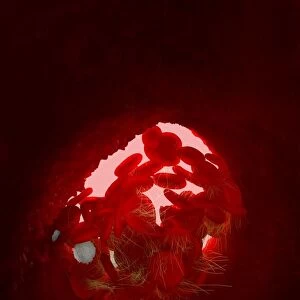
Cartilage cell, TEM C014 / 1434
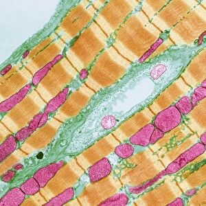
Cartilage cell. Transmission electron micrograph (TEM) of a section through a chondrocyte cell from hyaline cartilage of the trachea (windpipe). The cell nucleus (red) can be seen along with a large fat droplet (round, bright green) and glycogen granules (yellow). Chondrocytes are the only cells found in cartilage. They produce and maintain the cartilages extracellular matrix and. Hyaline cartilage is found in the epiphyseal plates of growing bones, and in the nose, larynx (voice box), trachea (windpipe) and bronchus. Magnification: x4000 when printed 10 centimetres wide
Science Photo Library features Science and Medical images including photos and illustrations
Media ID 9226817
© STEVE GSCHMEISSNER/SCIENCE PHOTO LIBRARY
Cartilage Cell Biology Chondrocyte Connective Tissue Cytological Cytology Extracellular Matrix Granules Histological Histology Hyaline Nucleus Trachea Transmission Electron Micrograph Transmission Electron Microscope Windpipe Section Sectioned
10"x8" Photo Print
Discover the intricacies of life with Media Storehouse's Photographic Prints. This captivating image, "Cartilage cell, TEM C014 / 1434" by STEVE GSCHMEISSNER/SCIENCE PHOTO LIBRARY, offers a fascinating glimpse into the world of science. Transmission Electron Micrograph (TEM) of a chondrocyte cell from hyaline cartilage of the trachea (windpipe) reveals the complex structure and functionality of this essential component of our bodies. Bring this mesmerizing piece of science art into your home or office and ignite curiosity and conversation. Media Storehouse's high-quality prints are perfect for laboratories, educational institutions, and anyone with an appreciation for the wonders of the natural world.
Photo prints are produced on Kodak professional photo paper resulting in timeless and breath-taking prints which are also ideal for framing. The colors produced are rich and vivid, with accurate blacks and pristine whites, resulting in prints that are truly timeless and magnificent. Whether you're looking to display your prints in your home, office, or gallery, our range of photographic prints are sure to impress. Dimensions refers to the size of the paper in inches.
Our Photo Prints are in a large range of sizes and are printed on Archival Quality Paper for excellent colour reproduction and longevity. They are ideal for framing (our Framed Prints use these) at a reasonable cost. Alternatives include cheaper Poster Prints and higher quality Fine Art Paper, the choice of which is largely dependant on your budget.
Estimated Product Size is 25.4cm x 20.3cm (10" x 8")
These are individually made so all sizes are approximate
Artwork printed orientated as per the preview above, with landscape (horizontal) orientation to match the source image.
EDITORS COMMENTS
This print showcases the intricate details of a cartilage cell, captured using a transmission electron microscope (TEM). The image depicts a section through a chondrocyte cell found in the hyaline cartilage of the trachea, also known as the windpipe. The focal point of this micrograph is the vibrant red nucleus of the chondrocyte, which stands out against its surroundings. Additionally, there is an intriguing presence of other cellular components within this tiny world. A large fat droplet can be observed as a round and bright green structure, adding to the complexity and diversity within this single cell. Furthermore, yellow glycogen granules are scattered throughout the image, providing insight into metabolic processes occurring within these specialized cells. Chondrocytes play a vital role in producing and maintaining the extracellular matrix that forms cartilage tissue. Hyaline cartilage primarily exists in various parts of our body such as epiphyseal plates in growing bones, nose, larynx (voice box), trachea (windpipe), and bronchus. This particular photograph has been magnified 4000 times to allow for detailed examination when printed at 10 centimeters wide. Steve Gschmeissner's expertise behind capturing this microscopic wonder from Science Photo Library offers us an awe-inspiring glimpse into the hidden beauty and complexity present at such minuscule scales.
MADE IN THE USA
Safe Shipping with 30 Day Money Back Guarantee
FREE PERSONALISATION*
We are proud to offer a range of customisation features including Personalised Captions, Color Filters and Picture Zoom Tools
SECURE PAYMENTS
We happily accept a wide range of payment options so you can pay for the things you need in the way that is most convenient for you
* Options may vary by product and licensing agreement. Zoomed Pictures can be adjusted in the Cart.