Photographic Print > Animals > Mammals > Muridae > Blue-grey Mouse
Photographic Print : Cerebellum tissue, light micrograph

Photo Prints from Science Photo Library
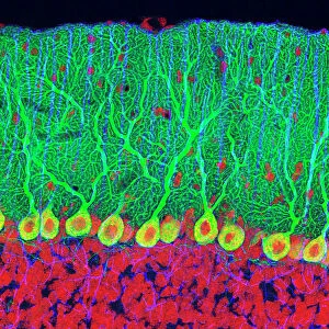
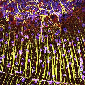
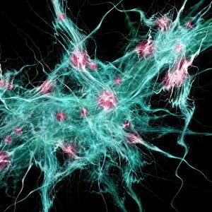
Cerebellum tissue, light micrograph
Cerebellum tissue. Confocal light micrograph of a section through the cerebellum of the brain. Purkinje cells, a type of neuron (nerve cell), are red. Glial cells, a type of support cell, are green, and cell nuclei are blue. Purkinje cells consist of a flask-shaped cell body with many branching processes (dendrites) that receive impulses from other cells. Purkinje cells form the junction between the granular and molecular layers of the grey matter of the cerebellum. The glial cells provide structural support, and nutrients and oxygen for the Purkinje cells. The cerebellum controls balance, posture and muscle coordination
Science Photo Library features Science and Medical images including photos and illustrations
Media ID 1697853
© C.J.GUERIN, PhD, MRC TOXICOLOGY UNIT/SCIENCE PHOTO LIBRARY
Central Nervous System Cerebellar Cerebellum Confocal Light Micrograph Dendrite Dendrites Fluorescence Fluorescent Glia Glial Cell Grey Matter Histological Histology Immunofluorescence Immunofluorescent Magnified Image Microscopic Subjects Nerve Cell Nervous Neuron Nuclei Nucleus Purkinje Cell Stain System Brain Cells Light Micrograph Light Microscope Neurological Neurology Section Sectioned
10"x10" Photo Print
Introducing the Media Storehouse range of Photographic Prints featuring the captivating image of "Cerebellum Tissue" by Science Photo Library. This stunning light micrograph offers a mesmerizing glimpse into the intricacies of the human body, showcasing a section through the cerebellum of the brain. Witness the vibrant colors of the Purkinje cells, a type of neuron, in brilliant red hues, contrasting against the intricate network of surrounding tissue. Bring the wonders of science into your home or office with this thought-provoking and visually striking piece. Order now and let this print ignite your curiosity and inspire your imagination.
Photo prints are produced on Kodak professional photo paper resulting in timeless and breath-taking prints which are also ideal for framing. The colors produced are rich and vivid, with accurate blacks and pristine whites, resulting in prints that are truly timeless and magnificent. Whether you're looking to display your prints in your home, office, or gallery, our range of photographic prints are sure to impress. Dimensions refers to the size of the paper in inches.
Our Photo Prints are in a large range of sizes and are printed on Archival Quality Paper for excellent colour reproduction and longevity. They are ideal for framing (our Framed Prints use these) at a reasonable cost. Alternatives include cheaper Poster Prints and higher quality Fine Art Paper, the choice of which is largely dependant on your budget.
Estimated Product Size is 25.4cm x 25.4cm (10" x 10")
These are individually made so all sizes are approximate
Artwork printed orientated as per the preview above, with landscape (horizontal) or portrait (vertical) orientation to match the source image.
FEATURES IN THESE COLLECTIONS
> Animals
> Mammals
> Muridae
> Blue-grey Mouse
> Posters
> Scientific Posters
EDITORS COMMENTS
This print showcases a confocal light micrograph of cerebellum tissue, providing us with a mesmerizing glimpse into the intricate world of the brain. The image reveals various components in vibrant colors - red representing Purkinje cells, green symbolizing glial cells, and blue indicating cell nuclei. Purkinje cells, characterized by their flask-shaped bodies and branching dendrites, play a crucial role in transmitting impulses from other cells within the cerebellum. Positioned at the junction between the granular and molecular layers of grey matter, these neurons are essential for maintaining balance, posture, and muscle coordination. Meanwhile, glial cells take on the responsibility of providing structural support to ensure optimal functioning of Purkinje cells. Additionally, they supply vital nutrients and oxygen necessary for neuronal health. Together with Purkinje cells' specialized functions and glial cell support system, this microscopic section illustrates how our central nervous system orchestrates complex biological processes. The magnified image offers an insight into neurology and histology while highlighting key elements such as dendrites and nuclei that contribute to overall brain function. This remarkable photograph not only captures scientific beauty but also serves as a reminder of our incredible human anatomy's intricacies.
MADE IN THE USA
Safe Shipping with 30 Day Money Back Guarantee
FREE PERSONALISATION*
We are proud to offer a range of customisation features including Personalised Captions, Color Filters and Picture Zoom Tools
SECURE PAYMENTS
We happily accept a wide range of payment options so you can pay for the things you need in the way that is most convenient for you
* Options may vary by product and licensing agreement. Zoomed Pictures can be adjusted in the Cart.