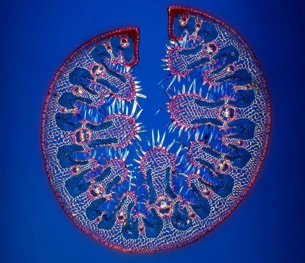
Ammophila arenaria leaf, light micrograph
![]()

Wall Art and Photo Gifts from Science Photo Library
Ammophila arenaria leaf, light micrograph
Ammophila arenaria leaf. Polarised light micrograph of a section through a marram grass (Ammophila arenaria) leaf, showing the characteristics that help reduce water loss. The outer epidermis (outer circle) consists of a layer of thick cuticle and layers of thick walled sclerenchyma (red), while the inner epidermis is folded and hairy (white) to trap water vapour. Chloroplasts (dark blue), phloem (dark blue) and xylem vessels (red) can also be seen. Magnification: x10 when printed 10 centimetres wide
Science Photo Library features Science and Medical images including photos and illustrations
Media ID 6353175
© DR KEITH WHEELER/SCIENCE PHOTO LIBRARY
Adaptation Adaptations Ammophila Arenaria Arid Adapted Bundles Cell Biology Chloroplast Chloroplasts Cuticle Cytological Cytology Epidermis Folded Hair Hairs Hairy Histological Histology Layer Marram Grass Microscopy Monocot Monocots Monocotyledon Monocotyledons Outer Epidermis Phloem Polarised Light Sclerenchyma Stain Stained Structural Structures Tissue Vascular Bundle Vessel Xerophyte Xerophytic Xylem Vessels Blue Background Cells Light Micrograph Light Microscope Section Sectioned
EDITORS COMMENTS
This print showcases the intricate details of an Ammophila arenaria leaf under a light microscope. The polarised light micrograph reveals the leaf's remarkable characteristics that aid in reducing water loss. The outer epidermis, depicted by the outer circle, is composed of a thick cuticle layer and layers of thick-walled sclerenchyma cells (shown in red). Meanwhile, the inner epidermis appears folded and hairy (depicted in white), serving as a clever mechanism to trap water vapor. Upon closer examination, one can observe chloroplasts (dark blue) responsible for photosynthesis, phloem vessels (dark blue) involved in nutrient transport, and xylem vessels (red) responsible for water transportation within the leaf structure. These components further highlight the complexity and functionality of this botanical wonder. The image's magnification allows for clear visibility when printed at 10 centimeters wide, enabling viewers to appreciate every minute detail. This photograph not only serves as an aesthetic masterpiece but also provides valuable insights into plant biology and adaptation strategies against arid environments. Captured by Science Photo Library's expert lens, this stunning micrograph offers a glimpse into nature's ingenuity while showcasing the beauty hidden within plants' cellular structures.
MADE IN THE USA
Safe Shipping with 30 Day Money Back Guarantee
FREE PERSONALISATION*
We are proud to offer a range of customisation features including Personalised Captions, Color Filters and Picture Zoom Tools
SECURE PAYMENTS
We happily accept a wide range of payment options so you can pay for the things you need in the way that is most convenient for you
* Options may vary by product and licensing agreement. Zoomed Pictures can be adjusted in the Cart.

