Capillary, TEM

![]()
Wall Art and Photo Gifts from Science Photo Library
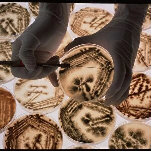
Capillary, TEM
Capillary. Transmission electron micrograph (TEM) of a section through a capillary, showing two red blood cells (erythrocytes, black) in its interior. Capillaries are the smallest diameter blood vessels (5-8 micrometres) and are just wide enough for red blood cells to pass along them. Their very thin walls are made of endothelial cells, across which gases, nutrients, fluids and cells may pass selectively in either direction. External to the endothelium, most capillaries have another layer of cells called pericytes that may be supportive (fibroblasts) or contractile (myofibroblasts). Magnification: x6, 000 when printed 10 centimetres wide
Science Photo Library features Science and Medical images including photos and illustrations
Media ID 9241349
© MICROSCAPE/SCIENCE PHOTO LIBRARY
Black And White Capillary Cell Biology Connective Tissue Contractile Cytological Cytology Endothelial Endothelium Erythrocyte Fibroblast Fibroblasts Histological Histology Layer Layers Lumen Organelle Organelles Red Blood Cell System Transmission Electron Micrograph Transmission Electron Microscope Vascular System Vessel Wall Blood Vessel Cells Circulatory System Section Sectioned
EDITORS COMMENTS
This print from Science Photo Library showcases the intricate structure of a capillary, one of the smallest blood vessels in our circulatory system. With a diameter ranging from 5 to 8 micrometers, capillaries provide just enough space for red blood cells to flow through their narrow lumen. The image reveals two erythrocytes, or red blood cells, gracefully gliding within the interior of the capillary. The thin walls of this microscopic vessel are composed of endothelial cells that allow selective passage of gases, nutrients, fluids, and even other cells in both directions. Beyond the endothelium lies another layer called pericytes. These specialized cells can either offer support as fibroblasts or exhibit contractile properties like myofibroblasts. Together with the endothelial layer, they contribute to maintaining the integrity and functionality of these vital structures within our body. With a magnification level of x6,000 when printed at 10 centimeters wide, this high-resolution transmission electron micrograph offers an extraordinary glimpse into cellular biology and histology. It highlights not only the complex organization but also emphasizes various organelles present within these tiny vessels. Science Photo Library continues to amaze us with its exceptional collection capturing fascinating aspects of biological systems such as this remarkable capillary sectioned under a transmission electron microscope.
MADE IN THE USA
Safe Shipping with 30 Day Money Back Guarantee
FREE PERSONALISATION*
We are proud to offer a range of customisation features including Personalised Captions, Color Filters and Picture Zoom Tools
SECURE PAYMENTS
We happily accept a wide range of payment options so you can pay for the things you need in the way that is most convenient for you
* Options may vary by product and licensing agreement. Zoomed Pictures can be adjusted in the Cart.