
Endothelium Collection
Endothelium: Unveiling the Intricate World of Blood Vessel Cells The endothelium, a fascinating cell structure that lines the inner surface of blood vessels


For sale as Licensed Images
Choose your image, Select your licence and Download the media
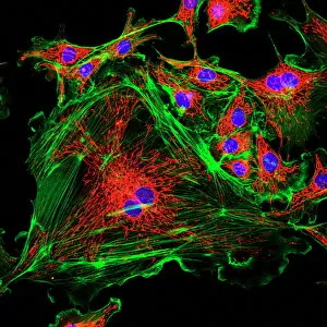
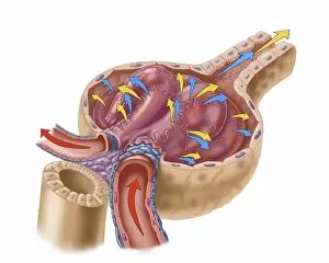
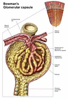
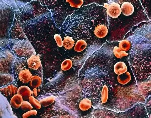
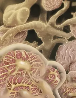
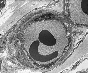
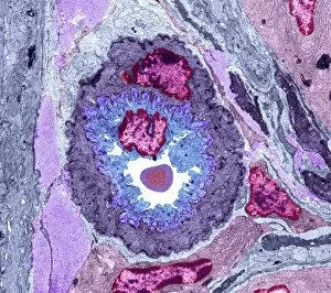
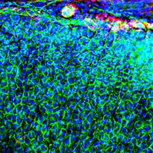
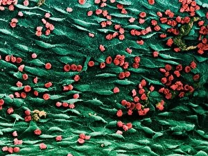
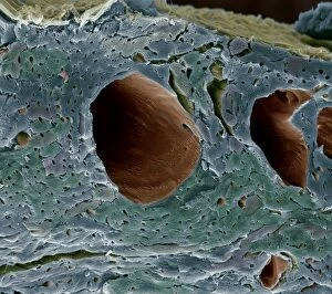
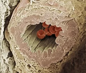
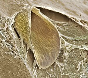
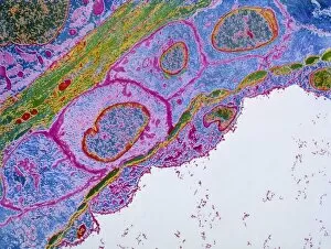
Endothelium: Unveiling the Intricate World of Blood Vessel Cells The endothelium, a fascinating cell structure that lines the inner surface of blood vessels, plays a crucial role in maintaining vascular health. Through its intricate network, it orchestrates various physiological processes and ensures proper functioning of vital organs. In a mesmerizing false-colour scanning electron microscope (SEM) image, we witness blood cells gracefully gliding along the endocardium – one layer of the endothelium found in the heart. This captivating sight reminds us of the ceaseless circulation within our bodies. An artist's depiction transports us to another realm – glomerulus capillaries within our kidneys. These tiny structures filter waste products from our blood and regulate fluid balance. The renal glomerulus is truly an architectural marvel designed for efficient filtration. Zooming further into this microscopic world, we explore the anatomy of Bowman's glomerular capsule – an essential component of each renal corpuscle. It envelops and protects delicate capillaries while facilitating selective filtration through its specialized membrane. As we delve deeper into understanding blood vessel anatomy, we are awestruck by its complexity and diversity throughout our body. From arteries to veins, these conduits transport oxygen-rich blood away from or towards our hearts with remarkable precision. Returning to the renal corpuscle and its filtration membrane, we appreciate how this dynamic duo maintains homeostasis by selectively allowing substances like water and electrolytes to pass through while retaining vital proteins and red blood cells within circulation. A closer look at a capillary alongside a red blood cell under transmission electron microscopy (TEM) reveals their intimate relationship - where nutrients are exchanged between circulating cells and tissues they nourish. This symbiotic dance sustains life itself. The endothelium unravels before us as an intricate tapestry interwoven with countless cellular threads that orchestrate essential functions within our bodies' vast network of vessels.