
Tendons Collection
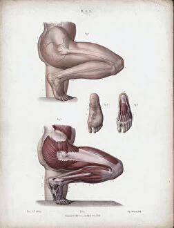
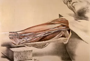
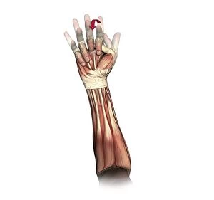
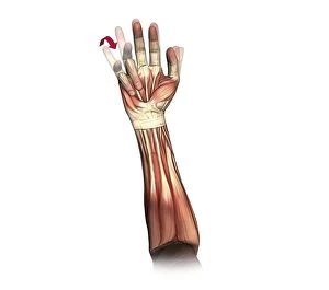
Tendons: The Unsung Heroes of the Muscular System From the intricate muscles on our head to the detailed foot anatomy depicted in a 19th-century illustration


For sale as Licensed Images
Choose your image, Select your licence and Download the media
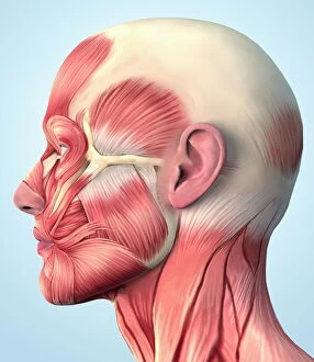
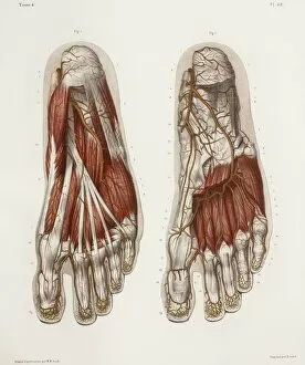
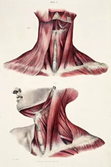
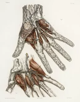
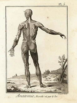
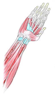
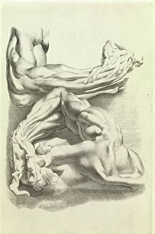
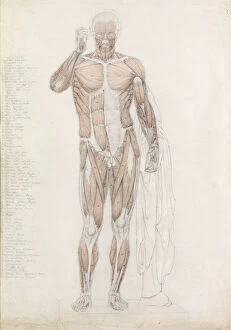
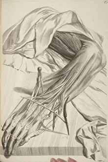
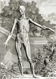
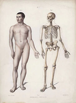
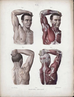
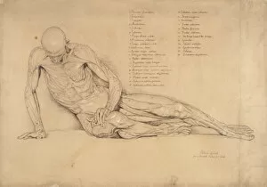
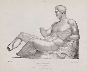
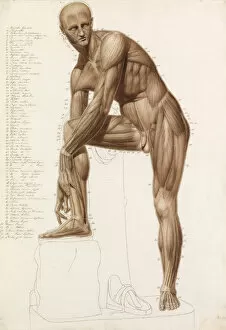
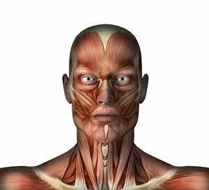
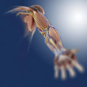
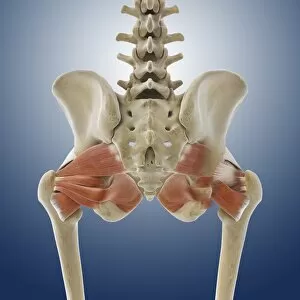
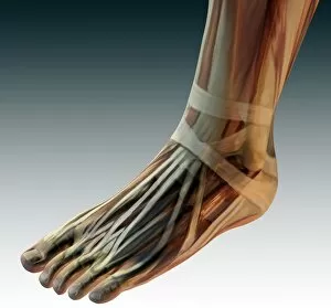
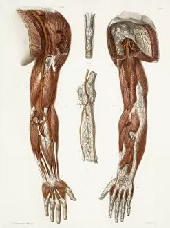
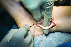
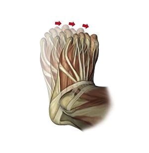
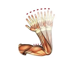
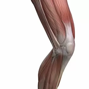
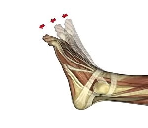
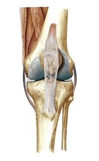
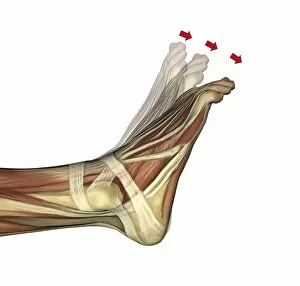
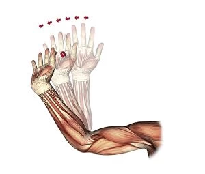
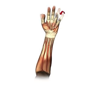
Tendons: The Unsung Heroes of the Muscular System From the intricate muscles on our head to the detailed foot anatomy depicted in a 19th-century illustration, tendons play a crucial role in connecting and supporting our body's musculature. These fibrous tissues are like bridges, linking muscles to bones and allowing us to move with grace and precision. In awe-inspiring artwork showcasing the muscles of the neck or female musculature, we witness how tendons seamlessly integrate into these complex systems. They act as strong cords that transmit force from contracting muscles, enabling fluid movements and maintaining stability. Shoulder muscles come alive in captivating artwork, highlighting their interplay with tendons. These resilient connective tissues anchor these powerful muscle groups, facilitating actions like lifting or throwing objects effortlessly. Delicate hand anatomy is beautifully portrayed through various artistic renditions. Tendons intricately weave through this masterpiece of dexterity, enabling precise finger movements necessary for tasks ranging from writing to creating art. Even Leonardo da Vinci recognized their significance centuries ago when he meticulously studied human anatomy. His drawings depict tendons alongside muscular structures, revealing his deep understanding of their vital role in movement. Moving down to the leg and foot region, we encounter yet another marvel - an illustration capturing the complexity of muscles and tendons working together harmoniously, and is here that we truly appreciate how tendons enable us to walk, run or dance by transmitting forces generated by our leg muscles onto our feet's skeletal framework. An enchanting study of human arm anatomy showcases both prominent biceps and triceps but also highlights lesser-known forearm and hand tendon networks. These often-overlooked heroes ensure smooth coordination between fingers' flexion and extension while gripping objects tightly or performing delicate tasks requiring fine motor skills. Lastly, an engraving depicting various muscle groups reminds us once again of tendons' indispensable presence within each one. Their strength allows us not only physical prowess but also the ability to express ourselves through movement.