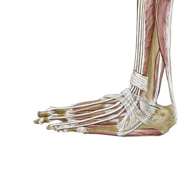
Foot ligaments and tendons, artwork C016 / 7010
![]()

Wall Art and Photo Gifts from Science Photo Library
Foot ligaments and tendons, artwork C016 / 7010
Foot ligaments and tendons. Artwork of the bones, ligaments (white, between bones), and tendons (white, muscle-bone) of the foot and the ankle joint from the side. This is a lateral view of the outside of the foot. The bones include the heel bone (calcaneus, lower right), the metatarsals (lower centre), and the phalanges (toe bones). The lower leg bones (upper right) are the tibia (covered in muscle, red) and the fibula. Major ligaments include the transverse crural and cruciate crural (straps). The extensor tendons connect lower leg muscles to the toes. The achilles tendon is at far right. For a sequence showing foot anatomy, see C016/7008 to C016/7010
Science Photo Library features Science and Medical images including photos and illustrations
Media ID 9246097
© D & L GRAPHICS / SCIENCE PHOTO LIBRARY
Achilles Tendon Ankle Bones Calcaneus Calf Muscle Cartilage Extensor Digitorum Brevis Extensor Hallucis Brevis Fibula Foot Gastrocnemius Heel Bone Joint Joints Lateral Ligament Ligaments Limb Metatarsal Metatarsals Peroneus Longus Phalanges Soleus Talus Tarsus Tibia Fibularis Longus Plantaris
EDITORS COMMENTS
This print showcases the intricate network of foot ligaments and tendons, providing a detailed glimpse into the complexity of our lower limb anatomy. Presented in a lateral view from the outside, this artwork highlights various key structures that contribute to our ability to walk, run, and maintain balance. The bones featured in this image include the calcaneus (heel bone), metatarsals (toe bones), and phalanges. Adjacent to these skeletal elements are major ligaments such as the transverse crural and cruciate crural, resembling sturdy straps that connect different parts of the foot together. The white ligaments can be seen delicately bridging between bones while ensuring stability during movement. Tendons play a crucial role in transmitting forces generated by muscles to produce motion. In this illustration, white tendons originating from lower leg muscles extend towards the toes via extensor tendons. Of particular significance is the prominent Achilles tendon on the far right side, connecting calf muscles to heel bone. With its clean white background contrasting against vibrant red muscle covering portions of tibia and fibula (lower leg bones), this artwork beautifully captures both scientific accuracy and artistic appeal. It serves as an invaluable resource for studying foot anatomy or understanding conditions related to joints, ligaments, or tendons within this region. Created by D & L GRAPHICS for Science Photo Library's extensive collection of scientific imagery, this visually striking print offers a fascinating insight into one's own body while celebrating human biology at its finest.
MADE IN THE USA
Safe Shipping with 30 Day Money Back Guarantee
FREE PERSONALISATION*
We are proud to offer a range of customisation features including Personalised Captions, Color Filters and Picture Zoom Tools
SECURE PAYMENTS
We happily accept a wide range of payment options so you can pay for the things you need in the way that is most convenient for you
* Options may vary by product and licensing agreement. Zoomed Pictures can be adjusted in the Cart.

