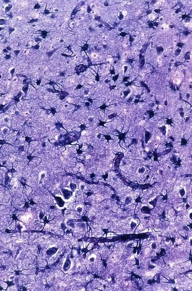
Glial cells, light micrograph
![]()

Wall Art and Photo Gifts from Science Photo Library
Glial cells, light micrograph
Glial cells in the brain. Light micrograph of a section through glial cells (dark) in the grey matter of the brain. Due to their star shape these glial cells are called astrocytes. Astrocytes are classified into fibrous types (seen here) and protoplasmic types. Fibrous astrocytes are not neurons (nerve cells) but serve as supporting cells in brain tissue with a range of functions. The astrocytes shown here have cytoplasmic extensions that are closely associated with blood vessels. Their function is to form a seal around the vessels to selectively restrict the entry of substances into the brain thus contributing to the blood-brain barrier. The total population of glial cells in the brain outnumbers neurons by a factor of ten and it is these cells that6
Science Photo Library features Science and Medical images including photos and illustrations
Media ID 9246859
© MICROSCAPE/SCIENCE PHOTO LIBRARY
Astrocyte Astrocytes Cell Biology Cytological Cytology Cytoplasmic Extension Extensions Fibrous Glial Cell Grey Matter Histological Histology Neuroscience Restriction Seal Selective Stain Stained Supporting Supportive Tissue Tissue Vessels Blood Vessel Brain Cells Light Micrograph Light Microscope Restrict Restrictive Section Sectioned
EDITORS COMMENTS
This print showcases glial cells, specifically astrocytes, in the grey matter of the brain. The star-shaped astrocytes are classified into fibrous types, as depicted here. While they may not be nerve cells like neurons, these fibrous astrocytes play a crucial role in supporting brain tissue with their diverse functions. The image reveals cytoplasmic extensions of the astrocytes that closely interact with blood vessels. Their primary function is to form a seal around these vessels, selectively restricting the entry of substances into the brain. This remarkable mechanism contributes to maintaining the integrity of the blood-brain barrier. It is fascinating to note that glial cells outnumber neurons by tenfold within our brains. These unsung heroes perform essential tasks and provide support for proper neural functioning. This visually stunning micrograph offers a glimpse into the intricate world of glial cells and their vital contributions to our neurological health. It serves as a testament to how microscopic structures can hold immense significance in understanding human anatomy and physiology. Captured through advanced light microscopy techniques, this histological image represents an amalgamation of biology and artistry. It invites us to appreciate both the complexity and beauty found within our own bodies while highlighting Science Photo Library's commitment to providing exceptional scientific imagery for educational purposes.
MADE IN THE USA
Safe Shipping with 30 Day Money Back Guarantee
FREE PERSONALISATION*
We are proud to offer a range of customisation features including Personalised Captions, Color Filters and Picture Zoom Tools
SECURE PAYMENTS
We happily accept a wide range of payment options so you can pay for the things you need in the way that is most convenient for you
* Options may vary by product and licensing agreement. Zoomed Pictures can be adjusted in the Cart.

