
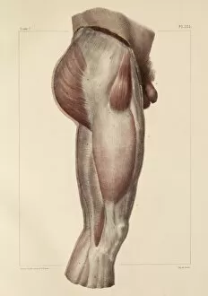
Fibrous Collection
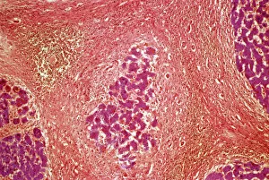
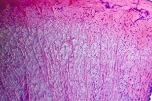
"Fibrous: Unveiling the Intricate Network of Connective Tissues" Liver tissue cirrhosis


For sale as Licensed Images
Choose your image, Select your licence and Download the media
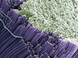
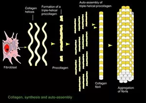
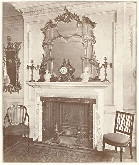
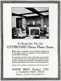
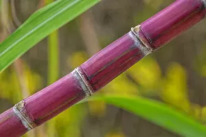
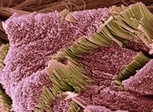
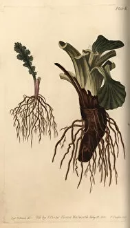
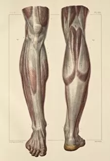
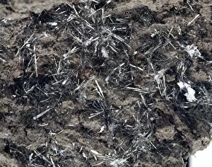
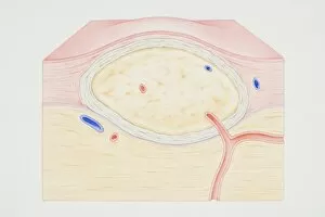
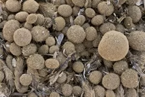
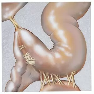
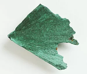
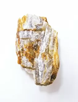
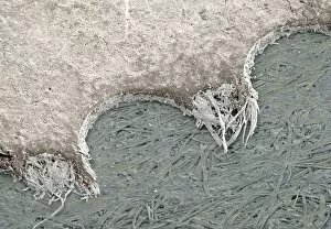
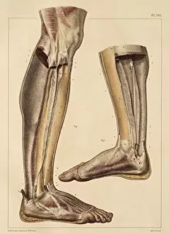
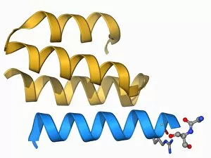
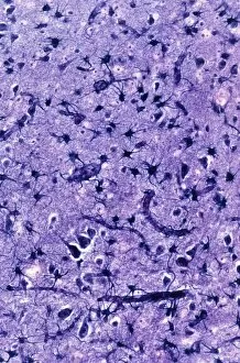
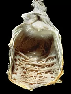
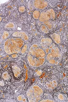
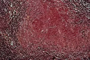
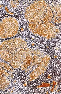
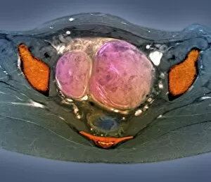
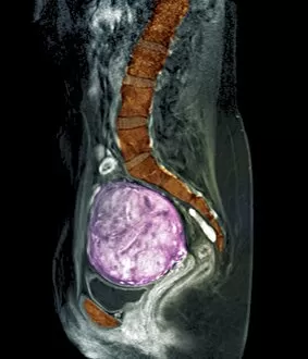
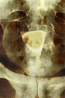
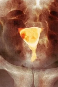
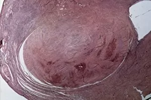
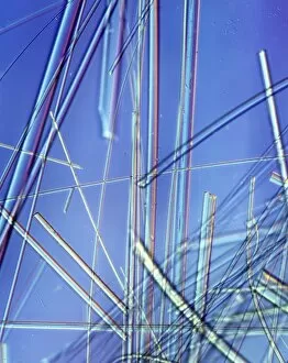
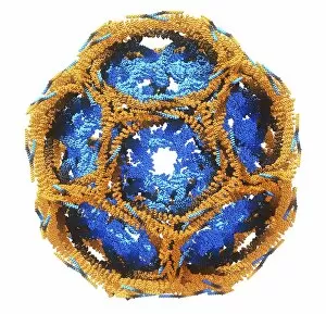
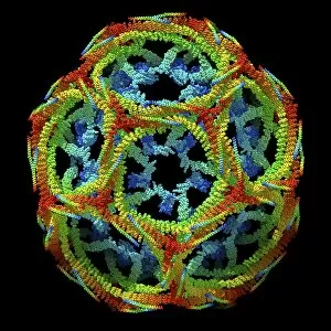
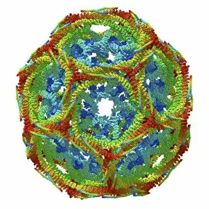
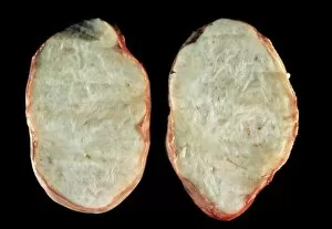
"Fibrous: Unveiling the Intricate Network of Connective Tissues" Liver tissue cirrhosis, light micrograph: A closer look at liver tissue reveals the devastating effects of cirrhosis, where fibrous bands disrupt its normal structure. Tendon, SEM: Under the scanning electron microscope, tendons appear as a dense and resilient fibrous matrix that enables our bodies to move with strength and precision. Collagen synthesis and assembly, artwork: Delicate brushstrokes depict the intricate process of collagen synthesis and assembly – a vital component in forming strong fibrous tissues throughout our body. J. S. Henry Ltd Fireplace and Surrounds: Craftsmanship meets elegance as this stunning fireplace surround showcases its ornate design featuring intricate fibrous patterns carved into high-quality materials. Noyes Bros Advertisement: Step into luxury with Noyes Bros' exquisite collection of furniture upholstered in sumptuous fabrics adorned with subtle yet striking fibrous motifs – an embodiment of refined taste. Plant roots: Fibrous root of groundsel Senecio - Explore nature's engineering marvels as we delve beneath the soil to uncover the complex network of fine fibrous roots supporting plants like groundsel Senecio. Sugar Cane: The tall stalks sway gracefully in fields filled with sugar cane – their tough but flexible fibers hold together this valuable crop that sweetens our lives. Tendon, SEM (again): Another glimpse through the lens reveals tendon fibers interwoven meticulously like a tightly-knit fabric; these resilient structures enable us to perform remarkable feats every day. Page from The Architects Compendium (litho): An architectural masterpiece comes alive on paper as detailed illustrations showcase how innovative designs incorporate both beauty and functionality using various fibrous materials. Page from The Architects Compendium (litho) [repeated].