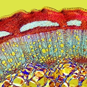
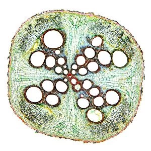
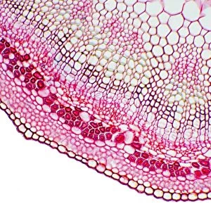
Pine needle, light micrograph

![]()
Wall Art and Photo Gifts from Science Photo Library
Pine needle, light micrograph
Pine needle. Light micrograph of a transverse section through a leaf (needle) of a pine tree (Pinus sp.). The leaves are needle-like in order to present a large surface area for photosynthesis but prevent too much water loss (transpiration). They have an epidermis (orange) of thick walled cells covered with a thick layer of cuticle. The mesophyll layer (green) under the epidermis is made up of parenchyma cells with in-folded walls, in-between these are resin canals (white circles). The vascular cylinder (centre) is surrounded by the endodermis (blue) which regulates water and mineral movement. Under the endodermis are two vascular bundles made up of phloem sieve cells (blue) and xylem tracheids (red). Magnification: x46 when printed 10 centimetres wide
Science Photo Library features Science and Medical images including photos and illustrations
Media ID 6299157
© DR KEITH WHEELER/SCIENCE PHOTO LIBRARY
Cell Biology Conifer Coniferous Cuticle Cytology Epidermis Gymnosperm Gymnosperms Needle Parenchyma Phloem Pine Plant Structure Stem Support Supportive Tracheid Tracheids Vascular Bundle Xylem Cells Light Micrograph Light Microscope Section Sectioned
MADE IN THE USA
Safe Shipping with 30 Day Money Back Guarantee
FREE PERSONALISATION*
We are proud to offer a range of customisation features including Personalised Captions, Color Filters and Picture Zoom Tools
SECURE PAYMENTS
We happily accept a wide range of payment options so you can pay for the things you need in the way that is most convenient for you
* Options may vary by product and licensing agreement. Zoomed Pictures can be adjusted in the Cart.