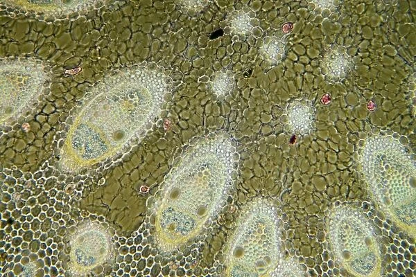
Sharp rush stem, light micrograph
![]()

Wall Art and Photo Gifts from Science Photo Library
Sharp rush stem, light micrograph
Sharp rush stem. Light micrograph of a section through the stem of a sharp rush (Juncus acutus) plant. This arid-adapted plant (xerophyte) has scattered vascular bundles. From the outer to the inner layer: thick-celled silicified epidermis (pink) covered with a thick cuticle (brown-green); triangular patches of fibres in-between which is columnar palisade tissue containing chloroplasts (blackish-brown); thick-walled parenchyma tissue (hexagonal); thin-walled parenchyma tissue containing oval vascular bundles; supporting fibres (whitish-pink); phloem (blue); xylem (pink-yellow). Magnification: x37 when printed 10 centimetres wide
Science Photo Library features Science and Medical images including photos and illustrations
Media ID 6353675
© DR KEITH WHEELER/SCIENCE PHOTO LIBRARY
Bundle Cell Biology Chloroplast Chloroplasts Columnar Cuticle Cytological Cytology Fibre Histological Histology Layer Layers Microscopy Monocot Monocots Monocotyledon Monocotyledons Parenchyma Phloem Stain Stained Stem Structural Structures Supporting Fibres Tissue Vascular Bundles Xerophyte Xylem Cells Light Micrograph Light Microscope Section Sectioned
EDITORS COMMENTS
This print showcases the intricate structure of a sharp rush stem, revealing its remarkable adaptations to arid environments. The light micrograph provides a detailed view of a cross-section through the stem, offering insights into the plant's cellular composition. The outermost layer is composed of thick-celled silicified epidermis, which appears in a striking pink hue and is protected by a robust brown-green cuticle. Moving inward, triangular patches of fibers can be observed interspersed with columnar palisade tissue containing chloroplasts that give off a blackish-brown coloration. Further inside, we encounter thick-walled parenchyma tissue arranged in hexagonal patterns. Within this layer lies thin-walled parenchyma tissue housing oval vascular bundles. Supporting fibers are also visible in whitish-pink shades. As we delve deeper into the stem's structure, distinct regions become apparent: phloem depicted in soothing blue tones and xylem exhibiting vibrant pink-yellow hues. This image not only highlights the complexity and diversity within plant tissues but also emphasizes how sharp rushes have evolved specialized features to thrive in harsh desert conditions as xerophytes. With scattered vascular bundles and an array of cell types strategically organized for optimal function, these plants exemplify nature's ingenuity at adapting to challenging environments. Captured at 37 times magnification when printed 10 centimeters wide, this stunning micrograph invites us to appreciate the hidden beauty found within botanical structures while shedding light on essential aspects of botany and biology.
MADE IN THE USA
Safe Shipping with 30 Day Money Back Guarantee
FREE PERSONALISATION*
We are proud to offer a range of customisation features including Personalised Captions, Color Filters and Picture Zoom Tools
SECURE PAYMENTS
We happily accept a wide range of payment options so you can pay for the things you need in the way that is most convenient for you
* Options may vary by product and licensing agreement. Zoomed Pictures can be adjusted in the Cart.

