Fine Art Print > Arts > Artists > G > Thomas Gray
Fine Art Print : Cerebellum structure, light micrograph


![]()
Fine Art Prints from Science Photo Library
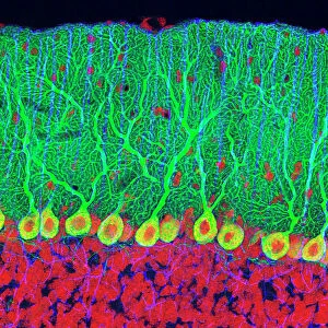
Cerebellum structure, light micrograph
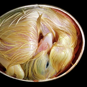
Cerebellum structure. Fluorescent light micrograph of a section through the cerebellum of the brain. The cerebellum comprises three main layers. The outer grey matter (cortex) comprises the molecular layer (green) and the granular layer (blue). The molecular layer is largely made up of the highly branched dendrites of Purkinje nerve cells (green), the large round bodies of which are found at the junction between the molecular and granular layers. Within the grey matter is the white matter (red), largely made up of the axons of the grey matters nerve cells. The cerebellum is involved in motor control, sensory perception and learning. Magnification: x20 when printed 10cm wide
Science Photo Library features Science and Medical images including photos and illustrations
Media ID 6421544
© THOMAS DEERINCK, NCMIR/SCIENCE PHOTO LIBRARY
Axon Axons Cerebellar Cerebellum Cortex Dendrite Dendrites Fluorescent Light Micrograph Granular Layer Gray Grey Matter Histological Histology Molecular Layer Nerve Cell Neuron Purkinje Cell White Matter Brain Light Microscope Nervous System Neurological Neurology
20"x20" (+3" Border) Fine Art Print
Discover the intricacies of the human body with our Fine Art Print from Media Storehouse's Science Collection. This captivating light micrograph of the Cerebellum structure, captured by Science Photo Library, offers a mesmerizing glimpse into the complex world of the brain. The cerebellum, a vital component of the nervous system, is depicted in stunning detail, revealing its three distinct layers. Bring this beautiful piece of scientific art into your home or office to inspire curiosity and ignite conversations about the wonders of the human body.
20x20 image printed on 26x26 Fine Art Rag Paper with 3" (76mm) white border. Our Fine Art Prints are printed on 300gsm 100% acid free, PH neutral paper with archival properties. This printing method is used by museums and art collections to exhibit photographs and art reproductions.
Our fine art prints are high-quality prints made using a paper called Photo Rag. This 100% cotton rag fibre paper is known for its exceptional image sharpness, rich colors, and high level of detail, making it a popular choice for professional photographers and artists. Photo rag paper is our clear recommendation for a fine art paper print. If you can afford to spend more on a higher quality paper, then Photo Rag is our clear recommendation for a fine art paper print.
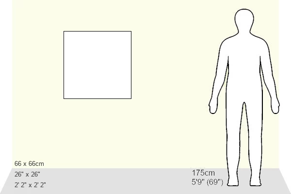
Estimated Image Size (if not cropped) is 50.8cm x 47.7cm (20" x 18.8")
Estimated Product Size is 66cm x 66cm (26" x 26")
These are individually made so all sizes are approximate
Artwork printed orientated as per the preview above, with landscape (horizontal) orientation to match the source image.
FEATURES IN THESE COLLECTIONS
> Animals
> Mammals
> Muridae
> Blue-grey Mouse
> Arts
> Artists
> G
> Thomas Gray
> Science Photo Library
> Specialist Imaging
EDITORS COMMENTS
This print showcases the intricate structure of the cerebellum, a vital part of the brain responsible for motor control, sensory perception, and learning. The image, captured using fluorescent light microscopy, reveals the complexity and beauty of this anatomical wonder. The cerebellum is composed of three main layers that can be distinguished in this micrograph. The outermost layer, known as the cortex or grey matter, consists of two distinct regions: the molecular layer (depicted in vibrant green) and the granular layer (displayed in striking blue). Within these layers lies an abundance of Purkinje nerve cells whose highly branched dendrites are represented by vivid green structures. These unique cells have round bodies located at the junction between the molecular and granular layers. Delving deeper into this mesmerizing composition reveals another crucial component: white matter depicted in intense red hues. Composed primarily of axons originating from nerve cells within grey matter areas, white matter plays a significant role in transmitting signals throughout different parts of our nervous system. This remarkable photograph not only captures scientific excellence but also serves as a testament to nature's artistry. Its detailed portrayal invites us to marvel at both its biological significance and aesthetic appeal.
MADE IN THE USA
Safe Shipping with 30 Day Money Back Guarantee
FREE PERSONALISATION*
We are proud to offer a range of customisation features including Personalised Captions, Color Filters and Picture Zoom Tools
SECURE PAYMENTS
We happily accept a wide range of payment options so you can pay for the things you need in the way that is most convenient for you
* Options may vary by product and licensing agreement. Zoomed Pictures can be adjusted in the Cart.