Framed Print > Science > SEM
Framed Print : Diatom alga, SEM


![]()
Framed Photos from Science Photo Library
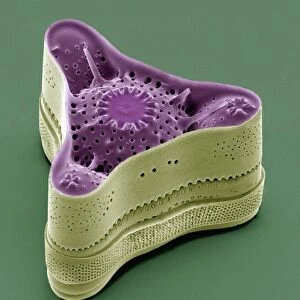
Diatom alga, SEM
Diatom. Coloured scanning electron micrograph (SEM) of the surface of the mineralised cell wall (frustule) of an unidentified diatom. This is a planktonic unicellular alga. The frustule contains silica and provides protection and support. Magnification unknown
Science Photo Library features Science and Medical images including photos and illustrations
Media ID 6292875
© STEVE GSCHMEISSNER/SCIENCE PHOTO LIBRARY
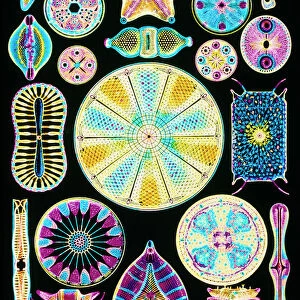
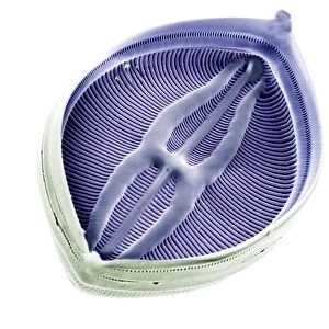
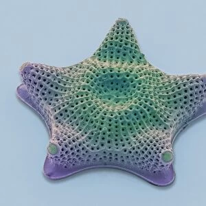
Alga Algae Algal Diatom Diatoms Frustule Honey Comb Lattice Net Work Phytoplankton Plankton Planktonic Siliceous Single Celled Surface Unicellular
12"x10" Modern Frame
Discover the intricacy of nature with our Media Storehouse Framed Prints featuring this captivating Coloured Scanning Electron Micrograph (SEM) of an unidentified Diatom alga by Science Photo Library. Witness the mineralised cell wall (frustule) of this planktonic unicellular alga in stunning detail. Each print is meticulously framed to showcase the beauty of this microscopic wonder, bringing a touch of the natural world into your home or office.
10x8 Print in an MDF Wooden Frame with 180 gsm Satin Finish Paper. Glazed using shatter proof thin plexi glass. Frame thickness is 1 inch and depth 0.75 inch. Fluted cardboard backing held with clips. Supplied ready to hang with sawtooth hanger and rubber bumpers. Spot clean with a damp cloth. Packaged foam wrapped in a card.
Contemporary Framed and Mounted Prints - Professionally Made and Ready to Hang
Estimated Image Size (if not cropped) is 25.4cm x 25.4cm (10" x 10")
Estimated Product Size is 25.4cm x 30.5cm (10" x 12")
These are individually made so all sizes are approximate
Artwork printed orientated as per the preview above, with landscape (horizontal) or portrait (vertical) orientation to match the source image.
EDITORS COMMENTS
This print showcases the intricate beauty of a Diatom alga, as captured by a scanning electron microscope (SEM). The image reveals the surface of the mineralized cell wall, known as the frustule, which provides both protection and support to this unidentified diatom. The vibrant colors in this photograph highlight the mesmerizing details of this planktonic unicellular alga. The frustule is composed mainly of silica, forming an enchanting honeycomb-like lattice structure that resembles a delicate network. This remarkable adaptation not only ensures stability for the diatom but also adds to its visual allure. With an unknown level of magnification employed in capturing this image, it becomes even more awe-inspiring to contemplate the hidden intricacies that exist within our natural world. This photograph serves as a reminder of nature's ability to create astonishingly complex forms on microscopic scales. Diatoms are part of the vast family of phytoplankton and play crucial roles in aquatic ecosystems. These single-celled organisms contribute significantly to global oxygen production and serve as essential food sources for various marine life forms. Science Photo Library has once again provided us with a stunning glimpse into nature's wonders through their exceptional photography skills. This particular print offers viewers an opportunity to appreciate both the scientific significance and aesthetic appeal found within these siliceous microorganisms without mentioning any commercial use associated with it.
MADE IN THE USA
Safe Shipping with 30 Day Money Back Guarantee
FREE PERSONALISATION*
We are proud to offer a range of customisation features including Personalised Captions, Color Filters and Picture Zoom Tools
SECURE PAYMENTS
We happily accept a wide range of payment options so you can pay for the things you need in the way that is most convenient for you
* Options may vary by product and licensing agreement. Zoomed Pictures can be adjusted in the Cart.