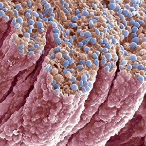
Framed Print > Animals > Fishes > G > Grouper
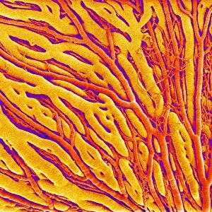
Framed Print : Eye muscle, SEM


![]()
Framed Photos from Science Photo Library
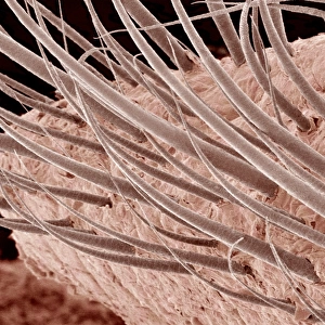
Eye muscle, SEM
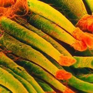
Eye muscle, coloured scanning electron micrograph (SEM). This is one of the rectus muscles of the eye. These muscles control the movement of the eyeball within the orbit (the eye socket). The strands are the muscle fibres that are grouped together to form the structure of the muscle. At top are strands of connective tissue
Science Photo Library features Science and Medical images including photos and illustrations
Media ID 6314563
© SUSUMU NISHINAGA/SCIENCE PHOTO LIBRARY
Bundle Bundles Colored Fibre Fibres Muscles Muscular Ocular Ophthalmology Tissue
12"x10" Modern Frame
Discover the intricacy of the human body with our Media Storehouse Framed Prints featuring the captivating image of "Eye muscle, SEM" by Science Photo Library. This mesmerizing coloured Scanning Electron Micrograph (SEM) reveals the intricate structure of one of the rectus muscles of the eye, showcasing its role in controlling the eyeball's movement within the orbit. Bring this stunning scientific image into your home or office as a conversation starter and a reminder of the marvels of the natural world. Our high-quality framed prints are meticulously crafted to preserve the rich details of the image and add a touch of sophistication to any space.
10x8 Print in an MDF Wooden Frame with 180 gsm Satin Finish Paper. Glazed using shatter proof thin plexi glass. Frame thickness is 1 inch and depth 0.75 inch. Fluted cardboard backing held with clips. Supplied ready to hang with sawtooth hanger and rubber bumpers. Spot clean with a damp cloth. Packaged foam wrapped in a card.
Contemporary Framed and Mounted Prints - Professionally Made and Ready to Hang
Estimated Image Size (if not cropped) is 25.4cm x 25.4cm (10" x 10")
Estimated Product Size is 30.5cm x 25.4cm (12" x 10")
These are individually made so all sizes are approximate
Artwork printed orientated as per the preview above, with landscape (horizontal) or portrait (vertical) orientation to match the source image.
FEATURES IN THESE COLLECTIONS
> Animals
> Fishes
> G
> Grouper
EDITORS COMMENTS
This print showcases the intricate beauty of an eye muscle, captured using a scanning electron microscope (SEM). The colored SEM image reveals one of the rectus muscles responsible for controlling the movement of the eyeball within its socket. These muscles play a crucial role in our ability to navigate and focus our vision. The mesmerizing strands depicted in this image are actually muscle fibers that come together to form the overall structure of this particular eye muscle. They appear like delicate threads woven with precision and purpose. At the top, we can observe strands of connective tissue that provide support and stability to ensure smooth functioning. This photograph not only highlights the anatomical details but also emphasizes how essential these muscles are for maintaining healthy ocular function. It serves as a reminder of just how intricately designed our bodies are, even at such microscopic levels. With its vibrant colors and stunning clarity, this print from Science Photo Library truly captures both the scientific significance and artistic appeal found within biology's hidden wonders. Whether you have an interest in anatomy or simply appreciate nature's marvels, this image is sure to captivate your imagination and spark curiosity about our incredible human physiology.
MADE IN THE USA
Safe Shipping with 30 Day Money Back Guarantee
FREE PERSONALISATION*
We are proud to offer a range of customisation features including Personalised Captions, Color Filters and Picture Zoom Tools
SECURE PAYMENTS
We happily accept a wide range of payment options so you can pay for the things you need in the way that is most convenient for you
* Options may vary by product and licensing agreement. Zoomed Pictures can be adjusted in the Cart.