
Activating Collection
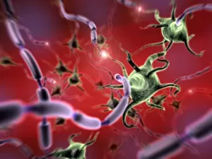
"Unlocking the Power Within: Activating Proteins and Gene Expression" In the intricate world of molecular biology


For sale as Licensed Images
Choose your image, Select your licence and Download the media
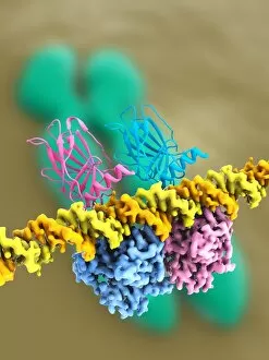
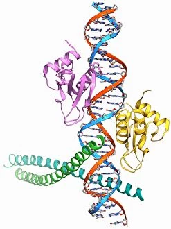
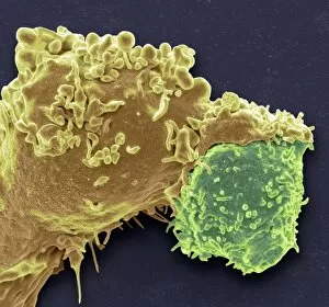
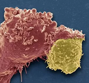
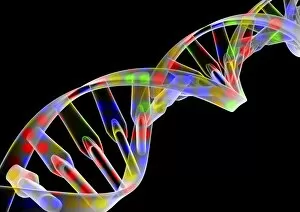
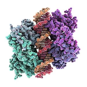
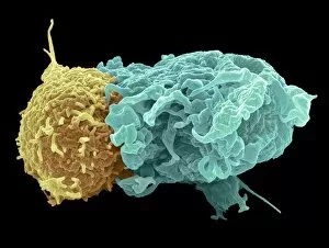
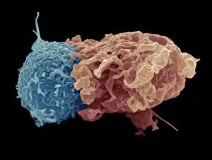
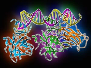
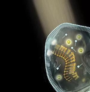
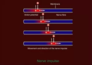
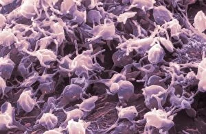
"Unlocking the Power Within: Activating Proteins and Gene Expression" In the intricate world of molecular biology, a fascinating process unfolds as proteins and genes are activated to carry out vital functions. Among these key players is the Ubiquitin activating enzyme protein E1 F007/9908, which initiates a cascade of events essential for cellular processes. Another crucial element in maintaining cellular integrity is the Tumour suppressor protein and DNA C017/3645. Its presence acts as a guardian against abnormal cell growth, ensuring our bodies remain healthy and free from potential threats. Visualizing these remarkable proteins becomes possible through detailed models that showcase their complex structures. One such model highlights the Tumour suppressor protein's intricate architecture, emphasizing its role in safeguarding our genetic material. Transcription activation of IFN-beta gene F006/9510 demonstrates how genes can be switched on to produce important molecules like interferons. This process plays a pivotal role in our immune response against infections and diseases. Gene activator proteins F006/9406 & F006/9269 further contribute to gene expression by binding to specific regions within DNA, kickstarting transcriptional machinery into action. Their involvement ensures that necessary instructions are followed accurately for proper cell function. White blood cells play an integral part in defending our bodies against foreign invaders. Antigen presentation by these cells (C016/9058 & C016/9057) allows them to communicate with other immune cells effectively, orchestrating an efficient defense strategy when faced with pathogens or cancerous cells. As we delve deeper into understanding these mechanisms, it becomes evident just how critical tumour suppressor proteins are for maintaining equilibrium within our bodies. Multiple molecular models emphasize their significance in preventing uncontrolled cell division – acting as powerful safeguards against cancer development. Through unraveling the mysteries behind activating proteins and genes, scientists inch closer towards harnessing this knowledge for medical advancements.