Alpha Helix Collection (#2)

The alpha helix, a fundamental structure in biology, plays a crucial role in various molecular processes

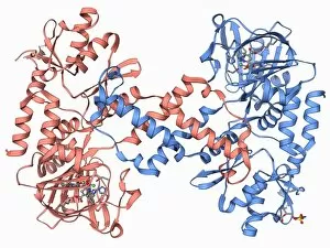
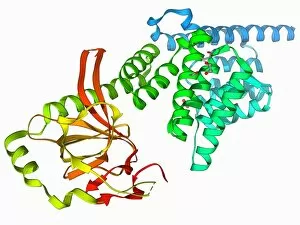
Background imageAlpha Helix Collection: Plexin signal transduction molecule F006 / 9751

Plexin signal transduction molecule F006 / 9751
Plexin signal transduction molecule. Molecular model of plexin-A4 a signal transduction protein that is involved in neural maintenance and regeneration

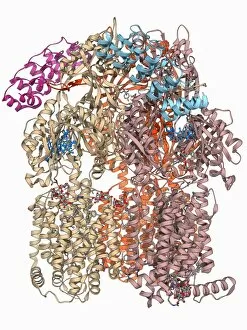
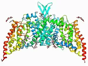
Background imageAlpha Helix Collection: Multidrug efflux pump molecule F006 / 9748

Multidrug efflux pump molecule F006 / 9748
Multidrug efflux pump. Molecular model of the multidrug efflux pump AcrB from the bacterium Escherichia coli transporting two doxorubicin molecules

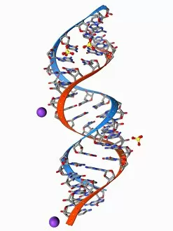
Background imageAlpha Helix Collection: RNA triplet repeat expansion F006 / 9749

RNA triplet repeat expansion F006 / 9749
RNA triplet repeat expansion. Molecular model of a CUG triplet repeat expansion in a molecule of double stranded RNA (ribonucleic acid)

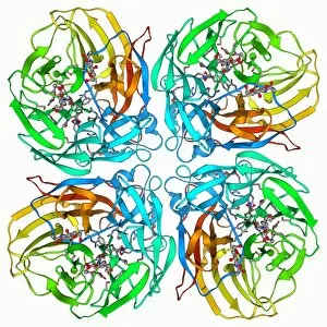
Background imageAlpha Helix Collection: Flu virus surface protein and drug F006 / 9745

Flu virus surface protein and drug F006 / 9745
Flu virus surface protein and drug. Molecular model of the neuraminidase glycoprotein enzyme from on the surface of the influenza A (flu) virus bound to the drug zanamivir

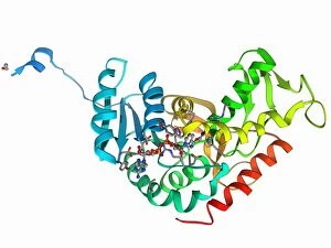
Background imageAlpha Helix Collection: Human lysine-specific demethylase F006 / 9747

Human lysine-specific demethylase F006 / 9747
Human lysine-specific demethylase, molecular model. This enzyme removes methyl groups from lysine residues in histones (proteins that package DNA)

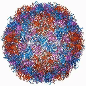
Background imageAlpha Helix Collection: Rhinovirus capsid, molecular model F006 / 9737

Rhinovirus capsid, molecular model F006 / 9737
Rhinovirus capsid, molecular model. This is human rhinovirus. The rhinovirus infects the upper respiratory tract and is the cause of the common cold. It is spread by coughs and sneezes
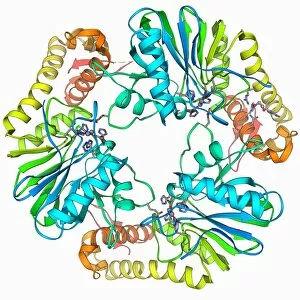
Background imageAlpha Helix Collection: Beta-lactamase molecule F006 / 9746

Beta-lactamase molecule F006 / 9746
Beta-lactamase enzyme, molecular model. This bacterial protein provides resistance to beta-lactam antibiotics, which include penicillin

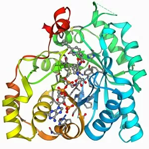
Background imageAlpha Helix Collection: Human muscle aldolase, molecular model F006 / 9742

Human muscle aldolase, molecular model F006 / 9742
Human muscle aldolase. Molecular model of the enzyme human muscle aldolase complexed with its substrate fructose 1, 6-bisphosphate

Background imageAlpha Helix Collection: ATP-binding cassette transporter F006 / 9743

ATP-binding cassette transporter F006 / 9743
ATP-binding cassette transporter. Molecular model of the human mitochondrial ATP-binding cassette transporter ABCB10. This protein is found on the inner membrane of mitochondria

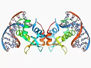
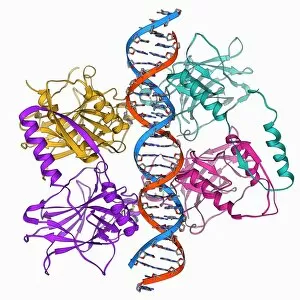
Background imageAlpha Helix Collection: Transcription factor bound to DNA F006 / 9744

Transcription factor bound to DNA F006 / 9744
Transcription factor bound to DNA. Molecular model of the human ETS translocation variant 1 (etv1) bound to a molecule of DNA (deoxyribonucleic acid)

Background imageAlpha Helix Collection: Beta-lactamase-like protein 2 molecule F006 / 9741

Beta-lactamase-like protein 2 molecule F006 / 9741
Beta-lactamase-like protein 2, molecular model

Background imageAlpha Helix Collection: Stress-responsive activator protein F006 / 9740

Stress-responsive activator protein F006 / 9740
Stress-responsive activator protein. Molecular model of the stress-responsive activator of p300 (strap) protein. This protein is activated when certain types of DNA (deoxyribonucleic acid)

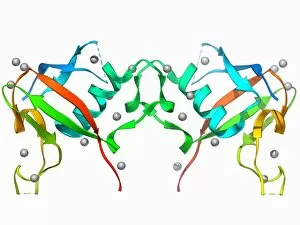
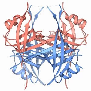
Background imageAlpha Helix Collection: Fatty acid binding protein 9 molecule F006 / 9739

Fatty acid binding protein 9 molecule F006 / 9739
Fatty acid binding protein 9. Molecular model of human testis-specific fatty acid binding protein 9

Background imageAlpha Helix Collection: Ebola virus glycoprotein and antibody F006 / 9738

Ebola virus glycoprotein and antibody F006 / 9738
Ebola virus glycoprotein and antibody. Molecular model of a surface glycoprotein from the Ebola virus (EBOV) bound to an antibody

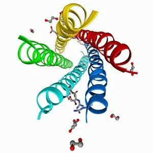
Background imageAlpha Helix Collection: Urea channel molecule F006 / 9735

Urea channel molecule F006 / 9735
Urea channel. Molecular model of the proton-gated urea channel from the bacterium Helicobacter pylori. This channel opens in acidic environments, allowing the entry of urea into the cell

Background imageAlpha Helix Collection: Leptin receptor molecule F006 / 9736

Leptin receptor molecule F006 / 9736
Leptin receptor. Molecular model of a leptin receptor complexed with an antibody. Leptin is a hormone produced by adipose (fat) tissue

Background imageAlpha Helix Collection: DNA clamp complexed with DNA molecule F006 / 9732

DNA clamp complexed with DNA molecule F006 / 9732
DNA clamp complexed with DNA molecule. Molecular model showing a sliding DNA (deoxyribonucleic acid) clamp (ring) complexed with a molecule of DNA (blue and red)

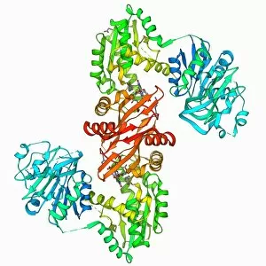
Background imageAlpha Helix Collection: GMP synthetase enzyme F006 / 9734

GMP synthetase enzyme F006 / 9734
GMP synthetase enzyme, molecular model. This enzyme, guanine monophosphate synthetase, catalyses the reaction that converts xanthosine monophosphate to guanosine monophosphate

Background imageAlpha Helix Collection: Single stranded DNA-binding protein F006 / 9733

Single stranded DNA-binding protein F006 / 9733
Single stranded DNA-binding protein (SSBP). Molecular model of a protein that binds to the single stranded DNA (deoxyribonucleic acid) in human mitochondria

Background imageAlpha Helix Collection: Sir3 gene silencer acting on DNA F006 / 9730

Sir3 gene silencer acting on DNA F006 / 9730
Sir3 gene silencer acting on DNA, molecular model. Sir3 (light blue) is acting on a circular strand of DNA (deoxyribonucleic acid, pink)

Background imageAlpha Helix Collection: Parathion hydrolase enzyme F006 / 9731

Parathion hydrolase enzyme F006 / 9731
Parathion hydrolase, molecular model. This enzyme hydrolyses bonds in organophosphates, which include pesticides and the nerve gas sarin

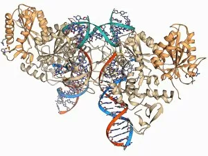
Background imageAlpha Helix Collection: Tumour suppressor protein with DNA F006 / 9729

Tumour suppressor protein with DNA F006 / 9729
Tumour suppressor protein. Molecular model of the tumour suppressor protein p53 (beige) bound to a molecule of DNA (deoxyribonucleic acid, red and blue)

Background imageAlpha Helix Collection: Iron-regulatory protein bound to RNA F006 / 9727

Iron-regulatory protein bound to RNA F006 / 9727
Iron-regulatory protein bound to RNA, molecular model. Iron regulatory protein 1 (IRP1, purple) bound to a short strand of RNA (ribonucleic acid, red) that includes iron-responsive elements (IREs)

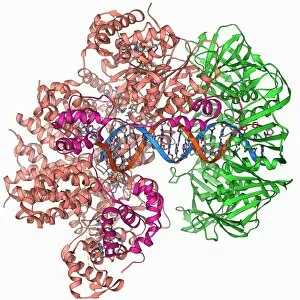
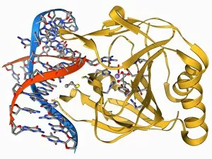
Background imageAlpha Helix Collection: DNA repair enzyme, molecular model F006 / 9726

DNA repair enzyme, molecular model F006 / 9726
DNA repair enzyme. Molecular model of the DNA (deoxyribonucleic acid) repair enzyme alpha-ketoglutarate-dependent dioxygenase bound to a molecule of DNA (red and blue)

Background imageAlpha Helix Collection: Oxidoreductase enzyme complex F006 / 9725

Oxidoreductase enzyme complex F006 / 9725
Oxidoreductase enzyme complex, molecular model. This is the membrane-bound domain formed from of a complex of NADH-quinone oxidoreductase subunits

Background imageAlpha Helix Collection: Saccharide transport protein F006 / 9722

Saccharide transport protein F006 / 9722
Saccharide transport protein. Molecular model of the phosphorylation-coupled saccharide transporter EIIC from the bacterium Bacillus cereus. EIIC is an integral membrane protein

Background imageAlpha Helix Collection: Aldo-keto reductase enzyme and ibuprofen F006 / 9724

Aldo-keto reductase enzyme and ibuprofen F006 / 9724
Aldo-keto reductase enzyme and ibuprofen. Molecular model of the enzyme aldo-keto reductase family 1 member C3 (AKR1C3) bound to a molecule of the anti-inflammatory drug ibuprofen

Background imageAlpha Helix Collection: SMAD4 protein domain bound to DNA F006 / 9723

SMAD4 protein domain bound to DNA F006 / 9723
SMAD4 protein domain bound to DNA, molecular model. This strand of DNA (deoxyribonucleic acid, red and blue) is surrounded by MH1 domains of the SMAD4 (Mothers against decapentaplegic homolog 4)

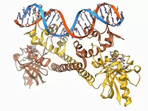
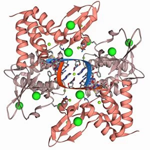
Background imageAlpha Helix Collection: Transcription activator and DNA F006 / 9721

Transcription activator and DNA F006 / 9721
Transcription activator and DNA. Molecular model of the transcriptional activator of the multidrug efflux transporter BmrR bound to a molecule of DNA (deoxyribonucleic acid, red and blue)

Background imageAlpha Helix Collection: Methyltransferase complexed with DNA F006 / 9711

Methyltransferase complexed with DNA F006 / 9711
Methyltransferase complexed with DNA, molecular model. The strand of DNA (deoxyribonucleic acid, red and blue) is enclosed by DNA methyltransferase 1 (DNMT-1, beige)

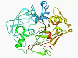
Background imageAlpha Helix Collection: Pepsinogen molecule F006 / 9710

Pepsinogen molecule F006 / 9710
Pepsinogen. Molecular model of pepsinogen, the inactive precursor to the digestive enzyme pepsin. Pepsion, which is released by the stomach digests proteins

Background imageAlpha Helix Collection: Glycosylation enzyme molecule F006 / 9708

Glycosylation enzyme molecule F006 / 9708
Glycosylation enzyme. Molecular model of the enzyme N-acetylglucosamine (GlcNAc) transferase. This intracellular enzyme adds N-acetylglucosamine molecules to target proteins

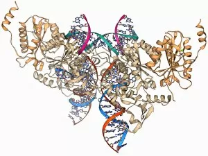
Background imageAlpha Helix Collection: Retroviral intasome molecule F006 / 9705

Retroviral intasome molecule F006 / 9705
Retroviral intasome molecule. Molecular model of an intasome from a retrovirus complexed with host cell DNA (deoxyribonucleic acid)

Background imageAlpha Helix Collection: Retroviral intasome molecule F006 / 9706

Retroviral intasome molecule F006 / 9706
Retroviral intasome molecule. Molecular model of an intasome from a retrovirus complexed with host cell DNA (deoxyribonucleic acid)

Background imageAlpha Helix Collection: Metarhodopsin molecule F006 / 9709

Metarhodopsin molecule F006 / 9709
Metarhodopsin II molecule, molecular model. This is one of the intermediary breakdown products formed when light falls on the photoreceptor pigment rhodopsin

Background imageAlpha Helix Collection: NpmA methyltransferase F006 / 9707

NpmA methyltransferase F006 / 9707
NpmA methyltransferase, molecular model. Methyltransferase enzymes act to add methyl groups to nucleic acids such as DNA (deoxyribonucleic acid), a process called DNA methylation

Background imageAlpha Helix Collection: Notch transcription complex F006 / 9703

Notch transcription complex F006 / 9703
Notch transcription complex. Molecular model of the notch transcription complex bound to the human Hes1 promoter on a strand of DNA (deoxyribonucleic acid, red and blue)

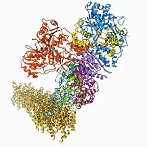
Background imageAlpha Helix Collection: Oxidoreductase enzyme complex F006 / 9700

Oxidoreductase enzyme complex F006 / 9700
Oxidoreductase enzyme complex. Molecular model of a complex of NADH-quinone oxidoreductase subunits. The whole is termed respiratory complex I

Background imageAlpha Helix Collection: DNA repair enzyme, molecular model F006 / 9704

DNA repair enzyme, molecular model F006 / 9704
DNA repair enzyme. Molecular model of the DNA (deoxyribonucleic acid) repair enzyme dioxygenase bound to a molecule of DNA (red and blue)

Background imageAlpha Helix Collection: Lassa virus nucleocapsid protein F006 / 9702

Lassa virus nucleocapsid protein F006 / 9702
Lassa virus nucleocapsid protein, molecular model. This protein has essential roles in RNA (ribonucleic acid) synthesis and immune suppression

Background imageAlpha Helix Collection: Human rotavirus enterotoxin F006 / 9701

Human rotavirus enterotoxin F006 / 9701
Human rotavirus enterotoxin. Molecular model of the NSP4 (nonstructural protein 4) from the human rotavirus. This enterotoxin induces diarrhoea

Background imageAlpha Helix Collection: Isocitrate dehydrogenase kinase F006 / 9698

Isocitrate dehydrogenase kinase F006 / 9698
Isocitrate dehydrogenase kinase. Molecular model of isocitrate dehydrogenase kinase phosphatase (AceK) complexed with its substrate isocitrate dehydrogenase (ICDH)

Background imageAlpha Helix Collection: Ebola viral protein 35 and RNA F006 / 9697

Ebola viral protein 35 and RNA F006 / 9697
Ebola viral protein 35 and RNA. Molecular model of the Ebola viral protein 35 (VP35) bound to a molecule of double stranded RNA (ribonucleic acid)

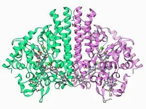
Background imageAlpha Helix Collection: Cytochrome C, molecular model F006 / 9696

Cytochrome C, molecular model F006 / 9696
Cytochrome C, molecular model. Cytochrome molecules perform oxidation and reduction reactions for electron transport, a chain of reactions used to power cellular processes that require energy

Background imageAlpha Helix Collection: Lactate dehydrogenase enzyme molecule F006 / 9699

Lactate dehydrogenase enzyme molecule F006 / 9699
Lactate dehydrogenase enzyme, molecular model. This enzyme converts pyruvate to lactate in the final steps of glycolysis, and lactate to pyruvate during the lactic acid cycle

Background imageAlpha Helix Collection: mRNA capping apparatus F006 / 9694

mRNA capping apparatus F006 / 9694
mRNA capping apparatus. Molecular model of the Cet-1-Ceg1 mRNA capping apparatus

Background imageAlpha Helix Collection: Eye lens protein molecule F006 / 9695

Eye lens protein molecule F006 / 9695
Eye lens protein. Molecular model of alpha-crystallin, a protein found in the lens of the eye. The regular arrangement of the protein in the lens is thought to be responsible for its transparency

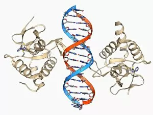
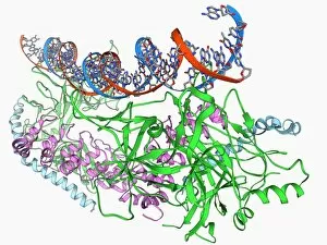
Background imageAlpha Helix Collection: Transcription repressor protein and DNA F006 / 9692

Transcription repressor protein and DNA F006 / 9692
Transcription repressor protein and DNA, molecular model. The repressor protein (green) is binding to a strand of DNA (deoxyribonucleic acid, pink and purple)


For sale as Licensed Images

Choose your image, Select your licence and Download the media

The alpha helix, a fundamental structure in biology, plays a crucial role in various molecular processes. From DNA transcription to protein synthesis, this intricate arrangement is found throughout the biological world. In the realm of genetics, the alpha helix participates in DNA transcription by aiding in the unwinding and separation of strands. Its elegant spiral shape allows for efficient reading and copying of genetic information. When it comes to proteins, the alpha helix serves as a secondary structure that contributes to their stability and function. Visualized through stunning artwork or molecular models, its coiled form adds strength and flexibility to these vital biomolecules. One example where we can observe this remarkable structure is within the nucleosome molecule. Here, DNA wraps around histone proteins forming tight coils resembling beads on a string – with each bead representing an alpha helix. Another instance occurs within bacterial ribosomes responsible for protein synthesis. The presence of multiple alpha helices enables precise positioning of molecules during translation – ensuring accurate assembly of amino acids into functional proteins. Viruses also exploit this structural motif; one such case being HIV reverse transcription enzyme. This enzyme utilizes an alpha helical region to convert viral RNA into DNA – a critical step in viral replication. Similarly, hepatitis C virus enzyme employs an intricate network of alpha helices depicted by molecular models. These structures aid in catalyzing chemical reactions necessary for viral survival and proliferation. Moving beyond viruses, manganese superoxide dismutase enzyme showcases how nature harnesses the power of the alpha helix for antioxidant defense mechanisms within cells. Its tightly wound coils protect against harmful free radicals that can damage cellular components. Alpha-helical motifs are not limited to enzymes alone but extend to larger molecules like human serum albumin or Argonaute protein involved in gene regulation pathways. Their well-defined arrangements contribute significantly to their respective functions within our bodies' complex systems.