Alpha Helix Collection (#5)

The alpha helix, a fundamental structure in biology, plays a crucial role in various molecular processes

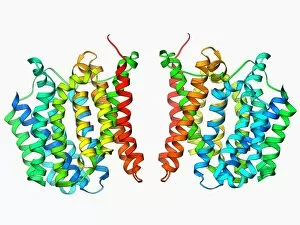
Background imageAlpha Helix Collection: Multidrug transporter molecule F006 / 9596

Multidrug transporter molecule F006 / 9596
Multidrug transporter. Molecular model of the multidrug transporter EmrD from the bacterium Escherichia coli. This protein pumps drugs, including antibiotics, out of the bacterial cell

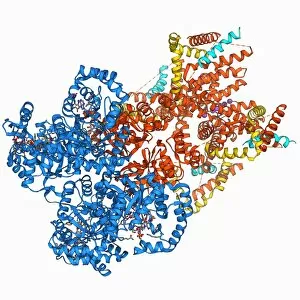
Background imageAlpha Helix Collection: Respiratory complex I F006 / 9593

Respiratory complex I F006 / 9593
Respiratory complex I. Molecular model of the hydrophilic domain of respiratory complex I from the bacterium Thermus thermophilus. This enzyme is involved in energy production

Background imageAlpha Helix Collection: Chymotrypsin digestive enzyme molecule F006 / 9577

Chymotrypsin digestive enzyme molecule F006 / 9577
Chymotrypsin digestive enzyme, molecular model. Chymotrypsin is a protease, an enzyme that breaks down proteins and peptides. It is secreted into the duodenum (small intestine) by the pancreas

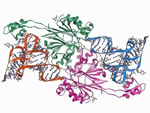
Background imageAlpha Helix Collection: Ribonuclease bound to transfer RNA F006 / 9591

Ribonuclease bound to transfer RNA F006 / 9591
Ribonuclease bound to transfer RNA, molecular model. This complex consists of the ribonuclease Z (RNase Z, green and pink) enzyme bound to a transfer RNA (tRNA) molecule (orange and blue)

Background imageAlpha Helix Collection: H5N1 Haemagglutinin protein subunit F006 / 9590

H5N1 Haemagglutinin protein subunit F006 / 9590
H5N1 haemagglutinin protein subunit. Molecular model of the haemagglutinin HA(5) subunit. Haemagglutinin is a surface protein from the influenza A virus

Background imageAlpha Helix Collection: RNA-induced silencing complex F006 / 9587

RNA-induced silencing complex F006 / 9587
RNA-induced silencing complex (RISC), molecular model. This complex consists of a bacterial argonaute protein (top right) bound to a small interfering RNA (siRNA) molecule (red and blue)

Background imageAlpha Helix Collection: RNA interference protein, molecular model F006 / 9589

RNA interference protein, molecular model F006 / 9589
RNA interference protein, molecular model. This RNA interference protein is also known as dicer. It is an RNAase enzyme that cleaves double-stranded RNA into short fragments called small interfering

Background imageAlpha Helix Collection: Acyl carrier protein molecule F006 / 9588

Acyl carrier protein molecule F006 / 9588
Acyl carrier protein (ACP), molecular model. This enzyme is involved in fatty acid synthesis

Background imageAlpha Helix Collection: DNA transcription, molecular model F006 / 9584

DNA transcription, molecular model F006 / 9584
DNA transcription. Molecular model of the enzyme RNA polymerase II synthesising a mRNA (messenger ribonucleic acid) strand from a DNA (deoxyribonucleic acid) template

Background imageAlpha Helix Collection: Dynamin enzyme, molecular model F006 / 9583

Dynamin enzyme, molecular model F006 / 9583
Dynamin enzyme. Molecular model of the pleckstrin homology (PH) domain of the dynamin enzyme. Domains are structural regions of enzymes that are often actively involved in biological processes

Background imageAlpha Helix Collection: Double-stranded RNA-ribonuclease III F006 / 9585

Double-stranded RNA-ribonuclease III F006 / 9585
Double-stranded RNA-ribonuclease III. Molecular model of ribonuclease III (RNase III, D44N, pink and green) complexed with a double-stranded RNA (ribonucleic acid) strand (red and blue)

Background imageAlpha Helix Collection: Human rotavirus capsid protein molecule F006 / 9582

Human rotavirus capsid protein molecule F006 / 9582
Human rotavirus capsid protein, molecular model

Background imageAlpha Helix Collection: Insulin receptor molecule F006 / 9581

Insulin receptor molecule F006 / 9581
Insulin receptor, molecular model. The insulin receptor is a transmembrane protein, that is it spans the cellular membrane

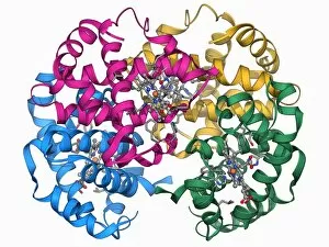
Background imageAlpha Helix Collection: Haemoglobin, molecular model F006 / 9580

Haemoglobin, molecular model F006 / 9580
Haemoglobin, molecular model. This is deoxyhaemoglobin, the molecule in its non-oxygen bound state. Haemoglobin transports oxygen around the body in red blood cells

Background imageAlpha Helix Collection: Bioluminescent enzyme molecule F006 / 9579

Bioluminescent enzyme molecule F006 / 9579
Bioluminescent enzyme. Molecular model of the bioluminescent enzyme luciferase from the Japanese aquatic firefly (Luciola cruciata) complexed with an intermediate protein

Background imageAlpha Helix Collection: Adenovirus penton base protein F006 / 9572

Adenovirus penton base protein F006 / 9572
Adenovirus penton base protein, molecular model. This protein molecule is a subunit called a penton, forming the vertices of the capsid of this adenovirus

Background imageAlpha Helix Collection: Fatty acid synthase molecule F006 / 9575

Fatty acid synthase molecule F006 / 9575
Fatty acid synthase (FAS), molecular model. FAS is a multi-enzyme that plays a key role in the synthesis of fatty acids (lipids) in the human body. It is not a single enzyme but a whole enzyme system

Background imageAlpha Helix Collection: Heat shock protein 90 chaperone complex F006 / 9576

Heat shock protein 90 chaperone complex F006 / 9576
Heat shock protein 90 (Hsp90) chaperone complex, molecular model. Hsp90 is a molecular chaperone and a member of the heat shock protein (HSP) family

Background imageAlpha Helix Collection: Chymotrypsin inhibitor 2 molecule F006 / 9578

Chymotrypsin inhibitor 2 molecule F006 / 9578
Chymotrypsin inhibitor 2, molecular model. This enzyme is a serine protease inhibitor (serpin) found in barley (Hordeum vulgare) seeds

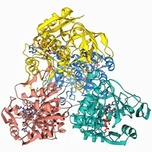
Background imageAlpha Helix Collection: Citrate synthase molecule F006 / 9573

Citrate synthase molecule F006 / 9573
Citrate synthase, molecular model. This enzyme is involved in the first step of the citric acid (or Krebs) cycle, the process by which mitochondria convert glucose to energy

Background imageAlpha Helix Collection: Family 84 glycoside hydrolase molecule F006 / 9574

Family 84 glycoside hydrolase molecule F006 / 9574
Family 84 glycoside hydrolase, molecular model. Glycoside hydrolases are enzymes that hydrolase the glycosidic bond between two or more carbohydrates

Background imageAlpha Helix Collection: Acetylcholine receptor molecule F006 / 9569

Acetylcholine receptor molecule F006 / 9569
Acetylcholine receptor. Molecular model showing the structure of a nicotinic acetlycholine receptor. This receptor, for the neurotransmitter acetylcholine

Background imageAlpha Helix Collection: Nuclear import complex molecule F006 / 9571

Nuclear import complex molecule F006 / 9571
Nuclear import complex. Molecular model of an importin protein complexed with a RanGTP molecule. This protein complex imports other proteins into the cell nucleus

Background imageAlpha Helix Collection: Retinal-producing oxygenase enzyme F006 / 9570

Retinal-producing oxygenase enzyme F006 / 9570
Retinal-producing oxygenase enzyme, molecular model. This enzyme plays a role in the production and metabolism of retinal and other apocarotenoids. Retinal is the chemical basis of vision in animals

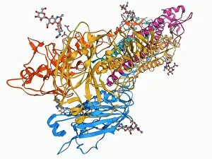
Background imageAlpha Helix Collection: Flu virus surface protein molecule F006 / 9566

Flu virus surface protein molecule F006 / 9566
Flu virus surface protein molecule. Molecular model of the neuraminidase glycoprotein enzyme found on the surface of the influenza A (flu) virus

Background imageAlpha Helix Collection: Amyloid beta protein molecule F006 / 9568

Amyloid beta protein molecule F006 / 9568
Amyloid beta protein. Molecular model showing the structure of amyloid beta protein, the primary component of amyloid plaques found in the brains of Alzheimers patients

Background imageAlpha Helix Collection: Calcium-binding protein molecule F006 / 9567

Calcium-binding protein molecule F006 / 9567
Calcium-binding protein. Molecule model of the calcium-binding protein calmodulin (CaM) bound to a myosin light-chain kinase molecule. This complex is involved in muscle contraction

Background imageAlpha Helix Collection: Iron-regulatory protein, molecular model F006 / 9565

Iron-regulatory protein, molecular model F006 / 9565
Iron-regulatory protein. Molecular model of iron regulatory protein 1 (IRP1). Depending on the conformation of IRP1, it can acts as either a regulator of mRNA (messenger RNA) or an enzyme

Background imageAlpha Helix Collection: Voltage-gated potassium channel F006 / 9562

Voltage-gated potassium channel F006 / 9562
Voltage-gated potassium (Kv) ion channel, molecular model. Ion channels are membrane-spanning proteins that form pores in cell membranes

Background imageAlpha Helix Collection: Tumour suppressor protein with DNA F006 / 9564

Tumour suppressor protein with DNA F006 / 9564
Tumour suppressor protein. Molecular model of the tumour suppressor protein p53 (beige) bound to a molecule of DNA (deoxyribonucleic acid, red and blue)

Background imageAlpha Helix Collection: Ricin molecule F006 / 9563

Ricin molecule F006 / 9563
Ricin. Molecular model of the toxic protein ricin. It comprises two entwined amino acid chains, termed A and B. The A-chain is toxic, inhibiting protein synthesis in cells

Background imageAlpha Helix Collection: DNA tetranucleosome, molecular model F006 / 9555

DNA tetranucleosome, molecular model F006 / 9555
DNA tetranucleosome. Molecular model of four nucleosomes, or a tetranucleosome. Nucleosomes are the fundamental repeating unit used to package DNA (deoxyribonucleic acid) inside cell nuclei

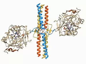
Background imageAlpha Helix Collection: Thrombin complexed with fibrinogen F006 / 9561

Thrombin complexed with fibrinogen F006 / 9561
Thrombin complexed with fibrinogen, molecular model. The thrombin molecules (beige, right and left) are bound to the central part of the fibrinogen molecule (down centre)

Background imageAlpha Helix Collection: SARS virus surface protein molecule F006 / 9560

SARS virus surface protein molecule F006 / 9560
SARS virus surface protein. Molecular model of the spike 2 (S2) envelope glycoprotein from the severe acute respiratory syndrome (SARS) virus

Background imageAlpha Helix Collection: DNA polymerase with DNA F006 / 9559

DNA polymerase with DNA F006 / 9559
DNA polymerase with DNA. Molecular model of human DNA polymerase beta (beige) complexed with a molecule of DNA (deoxyribonucleic acid, red and blue)

Background imageAlpha Helix Collection: Insulin molecule F006 / 9558

Insulin molecule F006 / 9558
Insulin, molecular model. Insulin plays an important role in blood sugar regulation. It is released from the pancreas when blood sugar levels are high, for example after a meal

Background imageAlpha Helix Collection: Bacteriophage DNA recombination F006 / 9554

Bacteriophage DNA recombination F006 / 9554
Bacteriophage DNA recombination. Molecular model showing DNA manipulation and recombination taking place at a Holliday junction with a bacteriophage enzyme

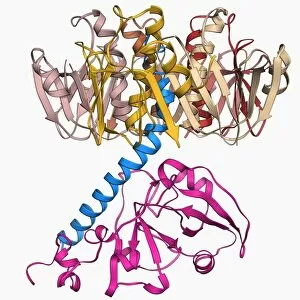
Background imageAlpha Helix Collection: ATP-dependent protease molecule F006 / 9552

ATP-dependent protease molecule F006 / 9552
ATP-dependent protease. Molecular model of the bacterial enzyme HsIUV protease. Proteases are enzymes that break down proteins. HsIUV is expressed in response to cellular stress

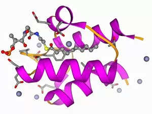
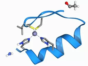
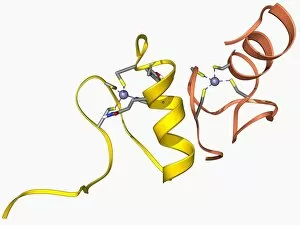
Background imageAlpha Helix Collection: Zinc finger molecule F006 / 9557

Zinc finger molecule F006 / 9557
Zinc finger, molecular model. Zinc fingers constitute the DNA recognition domains of many DNA regulatory proteins and are so named for their resemblance to fingers projecting from the protein

Background imageAlpha Helix Collection: Botulinum type B neurotoxin F006 / 9553

Botulinum type B neurotoxin F006 / 9553
Botulinum type B neurotoxin, molecular model. This powerful toxin, produced by the bacterium Clostridium botulinum, enters nerve cells and prevents release of neurotransmitters

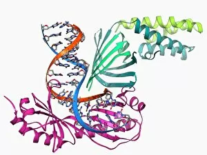
Background imageAlpha Helix Collection: TATA box-binding protein complex F006 / 9551

TATA box-binding protein complex F006 / 9551
TATA box-binding protein complex. Molecular model showing a yeast TATA box-binding protein (TBP) complexed with a strand of DNA (deoxyribonucleic acid, red and blue) and transcription factor IIA

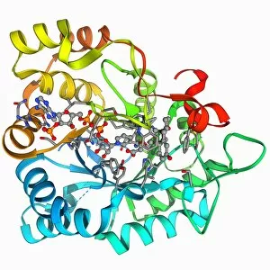
Background imageAlpha Helix Collection: NADP-dependent alcohol dehydrogenase F006 / 9549

NADP-dependent alcohol dehydrogenase F006 / 9549
NADP-dependent alcohol dehydrogenase, molecular model. Alcohol dehydrogenase (ADH) is an enzyme that facilitates the break-down of alcohols in the body, which could otherwise be toxic

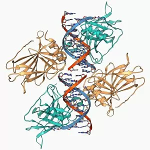
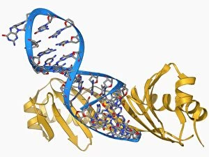
Background imageAlpha Helix Collection: TATA box-binding protein and DNA F006 / 9550

TATA box-binding protein and DNA F006 / 9550
TATA box-binding protein and DNA. Molecular model showing a TATA box-binding protein (TBP) complexed with a strand of DNA (deoxyribonucleic acid, red and blue)

Background imageAlpha Helix Collection: Cholera toxin molecule F006 / 9546

Cholera toxin molecule F006 / 9546
Cholera toxin, molecular model. Cholera toxin is a protein complex secreted by the bacterium Vibrio cholerae, that causes the disease cholera

Background imageAlpha Helix Collection: Programmed cell death protein 6 molecule F006 / 9548

Programmed cell death protein 6 molecule F006 / 9548
Programmed cell death protein 6, molecular model. This is a calcium-binding protein is involved in apoptosis (programmed cell death)

Background imageAlpha Helix Collection: GATA transcription factor and zinc finger F006 / 9547

GATA transcription factor and zinc finger F006 / 9547
GATA transcription factor. Molecular model of the GATA transcription factor bound to a zinc finger. Transcription factors are proteins that bind to specific DNA sequences

Background imageAlpha Helix Collection: Plant hormone regulator, molecular model F006 / 9545

Plant hormone regulator, molecular model F006 / 9545
Plant hormone regulator. Molecular model of Ia-amidohydrolase from thale cress (Arabidopsis thaliana). This enzyme acts to release the plant hormone indoe-3-acetic acid from its storage form

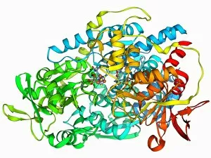
Background imageAlpha Helix Collection: Hydroxysteroid dehydrogenase molecule F006 / 9543

Hydroxysteroid dehydrogenase molecule F006 / 9543
Hydroxysteroid dehydrogenase. Molecular model of the human type 5 hydroxysteroid dehydrogenase enzyme bound to a molecule of the steroid drug androstene


For sale as Licensed Images

Choose your image, Select your licence and Download the media

The alpha helix, a fundamental structure in biology, plays a crucial role in various molecular processes. From DNA transcription to protein synthesis, this intricate arrangement is found throughout the biological world. In the realm of genetics, the alpha helix participates in DNA transcription by aiding in the unwinding and separation of strands. Its elegant spiral shape allows for efficient reading and copying of genetic information. When it comes to proteins, the alpha helix serves as a secondary structure that contributes to their stability and function. Visualized through stunning artwork or molecular models, its coiled form adds strength and flexibility to these vital biomolecules. One example where we can observe this remarkable structure is within the nucleosome molecule. Here, DNA wraps around histone proteins forming tight coils resembling beads on a string – with each bead representing an alpha helix. Another instance occurs within bacterial ribosomes responsible for protein synthesis. The presence of multiple alpha helices enables precise positioning of molecules during translation – ensuring accurate assembly of amino acids into functional proteins. Viruses also exploit this structural motif; one such case being HIV reverse transcription enzyme. This enzyme utilizes an alpha helical region to convert viral RNA into DNA – a critical step in viral replication. Similarly, hepatitis C virus enzyme employs an intricate network of alpha helices depicted by molecular models. These structures aid in catalyzing chemical reactions necessary for viral survival and proliferation. Moving beyond viruses, manganese superoxide dismutase enzyme showcases how nature harnesses the power of the alpha helix for antioxidant defense mechanisms within cells. Its tightly wound coils protect against harmful free radicals that can damage cellular components. Alpha-helical motifs are not limited to enzymes alone but extend to larger molecules like human serum albumin or Argonaute protein involved in gene regulation pathways. Their well-defined arrangements contribute significantly to their respective functions within our bodies' complex systems.