Alpha Helix Collection (#9)

The alpha helix, a fundamental structure in biology, plays a crucial role in various molecular processes

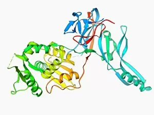
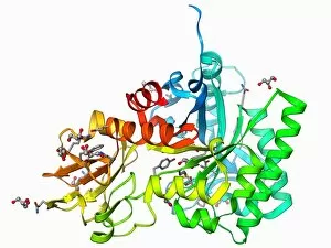
Background imageAlpha Helix Collection: Proteinase inhibitor molecule F006 / 9394

Proteinase inhibitor molecule F006 / 9394
Proteinase inhibitor. Molecular model of a proteinase inhibitor, or antitrypsin, molecule bound to a tryspin protease. The proteinase inhibitor is a type of serine protease inhibitor (serpin)

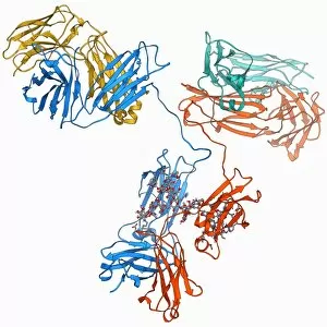
Background imageAlpha Helix Collection: Voltage-gated potassium channel F006 / 9391

Voltage-gated potassium channel F006 / 9391
Voltage-gated potassium channel. Molecular model of a voltage-gated potassium (Kv) ion channel complexed with the antigen-binding fragment (Fab) of a monoclonal antibody

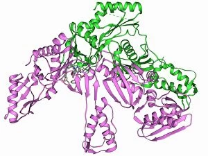
Background imageAlpha Helix Collection: DNA polymerase IV with DNA F006 / 9390

DNA polymerase IV with DNA F006 / 9390
polymerase IV with DNA. Molecular model of DNA polymerase IV (beige) complexed with a molecule of DNA (deoxyribonucleic acid, red and blue)

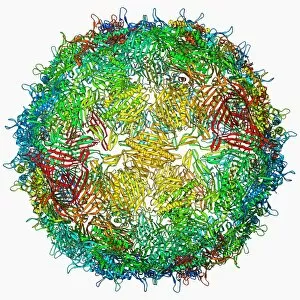
Background imageAlpha Helix Collection: Coxsackie B3 virus particle F006 / 9381

Coxsackie B3 virus particle F006 / 9381
Coxsackie B3 virus. Computer model of the capsid of the Coxsackie B3 virus
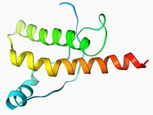
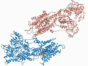
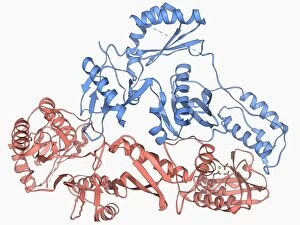
Background imageAlpha Helix Collection: Integrin, molecular model F006 / 9388

Integrin, molecular model F006 / 9388
Integrin. Molecular model of the integrin protein alpha-v beta-3. This is a transmembrane protein that is found on platelets. It is composed of two subunits; integrin alpha-V and integrin beta 3

Background imageAlpha Helix Collection: VMA-1 derived endonuclease molecule F006 / 9389

VMA-1 derived endonuclease molecule F006 / 9389
VMA-1 derived endonuclease. Molecular model of the VMA-1 derived endonuclease restriction enzyme. Restriction enzymes, also known as restriction endonucleases

Background imageAlpha Helix Collection: Uroporphyrinogen III decarboxylase F006 / 9387

Uroporphyrinogen III decarboxylase F006 / 9387
Uroporphyrinogen III decarboxylase. Molecular model of the enzyme human uroporphyrinogen III decarboxylase (UROD). Mutations or deficiencies in this enzyme cause the disorder porphyria with a build

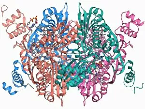
Background imageAlpha Helix Collection: HIV reverse transcription enzyme F006 / 9385

HIV reverse transcription enzyme F006 / 9385
HIV reverse transcription enzyme. Molecular model of the reverse transcriptase enzyme found in HIV (the human immunodeficiency virus) bound to the inhibitor nevirapine

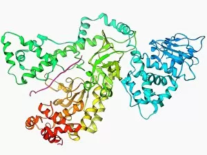
Background imageAlpha Helix Collection: Anthrax lethal factor molecule F006 / 9384

Anthrax lethal factor molecule F006 / 9384
Anthrax lethal factor, molecular model. This enzyme is one of three protein components that form the anthrax toxin produced by the bacterium Bacillus anthracis

Background imageAlpha Helix Collection: Early endosome antigen 1 molecule F006 / 9386

Early endosome antigen 1 molecule F006 / 9386
Early endosome antigen 1 (EEA1), molecular model. This protein is involved in the fusion and sorting of endosomes (membrane-bound compartments used to transport molecules)

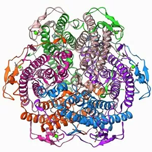
Background imageAlpha Helix Collection: Manganese catalase enzyme molecule F006 / 9383

Manganese catalase enzyme molecule F006 / 9383
Manganese catalase. Molecular model of the enzyme manganese catalase from the bacterium Lactobacillus plantarum. This enzyme catalyses the break down of hydrogen peroxide to water and oxygen

Background imageAlpha Helix Collection: Photosystem I molecule F006 / 9380

Photosystem I molecule F006 / 9380
Photosystem I. Molecular model of the photosystem I complex from the cyanobacterium Synechococcus elongatus. Shown here are beta-carotene, alpha-chlorophyll and reaction centre subunits

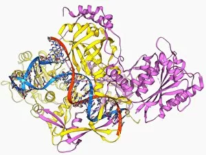
Background imageAlpha Helix Collection: Repair protein and DNA, molecular model F006 / 9382

Repair protein and DNA, molecular model F006 / 9382
Repair protein and DNA. Molecular model of the Ku heterodimer (yellow and pink) bound to a strand of DNA (deoxyribonucleic acid, red and blue) as part of the repair process

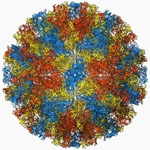
Background imageAlpha Helix Collection: Norwalk virus capsid, molecular model F006 / 9372

Norwalk virus capsid, molecular model F006 / 9372
Norwalk virus capsid, molecular model. This norovirus, which causes a viral form of gastroenteritis, is transmitted from person-to-person or through contaminated food

Background imageAlpha Helix Collection: Anthrax lethal factor molecule F006 / 9379

Anthrax lethal factor molecule F006 / 9379
Anthrax lethal factor, molecular model. This enzyme is one of three protein components that form the anthrax toxin produced by the bacterium Bacillus anthracis

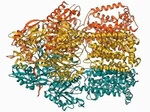
Background imageAlpha Helix Collection: Multidrug efflux pump molecule F006 / 9376

Multidrug efflux pump molecule F006 / 9376
Multidrug efflux pump. Molecular model of the multidrug efflux pump AcrB from the bacterium Escherichia coli. This protein pumps drugs, including antibiotics, out of the bacterial cell

Background imageAlpha Helix Collection: Calcium pumping ATPase muscle enzyme F006 / 9377

Calcium pumping ATPase muscle enzyme F006 / 9377
Calcium pumping ATPase enzyme. Molecular model of the ATPase enzyme that pumps calcium in and out of muscle cells and controls muscle contractions

Background imageAlpha Helix Collection: Epidermal growth factor and receptor F006 / 9375

Epidermal growth factor and receptor F006 / 9375
Epidermal growth factor molecule. Molecular model of epidermal growth factor (EGF) bound to a receptor. EGF plays an important role in the regulation of cell growth, proliferation and differentiation

Background imageAlpha Helix Collection: Amyloid beta protein molecule F006 / 9378

Amyloid beta protein molecule F006 / 9378
Amyloid beta protein. Molecular model showing the structure of amyloid beta protein, the primary component of amyloid plaques found in the brains of Alzheimers patients

Background imageAlpha Helix Collection: Chitinase enzyme molecule F006 / 9374

Chitinase enzyme molecule F006 / 9374
Chitinase, molecular model. This enzyme catalyses the break down of glycosidic bonds in chitin, the main component of fungal cell walls and the exoskeletons of arthropods

Background imageAlpha Helix Collection: Immunoglobulin G antibody molecule F006 / 9371

Immunoglobulin G antibody molecule F006 / 9371
Immunoglobulin G (IgG) antibody, molecular model. This is the most abundant immunoglobulin and is found in all body fluids

Background imageAlpha Helix Collection: Human interferon alpha molecule F006 / 9373

Human interferon alpha molecule F006 / 9373
Human interferon alpha, molecular model. Interferons are proteins produced by white blood cells as part of the immune response to invading pathogens, especially viruses

Background imageAlpha Helix Collection: DNA transcription, molecular model F006 / 9368

DNA transcription, molecular model F006 / 9368
DNA transcription. Molecular model of the enzyme RNA polymerase II synthesising a mRNA (messenger ribonucleic acid) strand from a DNA (deoxyribonucleic acid) template

Background imageAlpha Helix Collection: Hexokinase sugar enzyme F006 / 9370

Hexokinase sugar enzyme F006 / 9370
Hexokinase, molecular model. This enzyme promotes the conversion (phosphorylation) of glucose into glucose 6-phosphate. Cells then use the glucose 6-phosphate when they require energy

Background imageAlpha Helix Collection: Eye lens protein molecule F006 / 9367

Eye lens protein molecule F006 / 9367
Eye lens protein. Molecular model of delta-crystallin, a protein found in the lens of the eye. The regular arrangement of the protein in the lens is thought to be responsible for its transparency

Background imageAlpha Helix Collection: Cholesterol producing enzyme and statin F006 / 9366

Cholesterol producing enzyme and statin F006 / 9366
Cholesterol producing enzyme and statin. Computer model showing the molecular structure of HMG-CoA reductase (HMGCR) in complex with Atorvastatin

Background imageAlpha Helix Collection: Interleukin-1 beta molecule F006 / 9369

Interleukin-1 beta molecule F006 / 9369
Interleukin-1 beta, molecular model. This cytokine immune protein is an important mediator of the inflammatory response

Background imageAlpha Helix Collection: Cholesterol producing enzyme molecule F006 / 9365

Cholesterol producing enzyme molecule F006 / 9365
Cholesterol producing enzyme. Molecular model of HMG-CoA reductase (HMGCR), the rate-controlling enzyme of the metabolic pathway that produces cholesterol in the body

Background imageAlpha Helix Collection: Bacterial cell wall enzyme molecule F006 / 9364

Bacterial cell wall enzyme molecule F006 / 9364
Bacterial cell wall enzyme. Molecular model of D-alanyl-D-alanine carboxypeptidase, a transpeptidase. This enzyme cross-links peptidoglycan chains in bacterial cell walls, making them rigid

Background imageAlpha Helix Collection: Mitochondrial processing peptidase F006 / 9362

Mitochondrial processing peptidase F006 / 9362
Yeast mitochondrial processing peptidase, molecular model. This enzyme plays an essential role in importing mitochondrial proteins

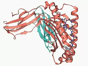
Background imageAlpha Helix Collection: MHC protein-antigen complex F006 / 9363

MHC protein-antigen complex F006 / 9363
MHC protein-antigen complex. Molecular model of the human class I MHC (major histocompatibility complex) protein HLA-B27 complexed with beta-2 microglobulin

Background imageAlpha Helix Collection: Phosphoglucose isomerase molecule F006 / 9361

Phosphoglucose isomerase molecule F006 / 9361
Phosphoglucose isomerase molecule. Molecular model of a rabbit phosphoglucose isomerase molecule complexed with a fructose-6-phosphate molecule

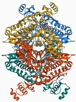
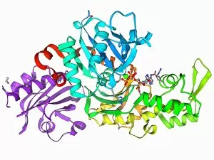
Background imageAlpha Helix Collection: HIV reverse transcription enzyme F006 / 9360

HIV reverse transcription enzyme F006 / 9360
HIV reverse transcription enzyme. Molecular model of the reverse transcriptase enzyme found in HIV (the human immunodeficiency virus)

Background imageAlpha Helix Collection: HGPRTase molecule F006 / 9359

HGPRTase molecule F006 / 9359
HGPRTase. Molecular model of hypoxanthine-guanine phosphoribosyltransferase (HGPRTase) bound to a molecule of guanosine monophosphate (GMP). HGPRTase is involved in the purine salvage pathway

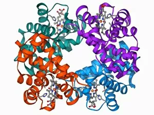
Background imageAlpha Helix Collection: Haemoglobin molecule F006 / 9356

Haemoglobin molecule F006 / 9356
Haemoglobin, molecular model. Haemoglobin is a metalloprotein that transports oxygen around the body in red blood cells. Each molecule consists of iron-containing haem groups (sticks)

Background imageAlpha Helix Collection: Beta-actin molecule F006 / 9358

Beta-actin molecule F006 / 9358
Beta-actin. Molecular model of the cytoskeletal protein beta-actin bound to profilin. The cytoskeleton maintains the cells shape

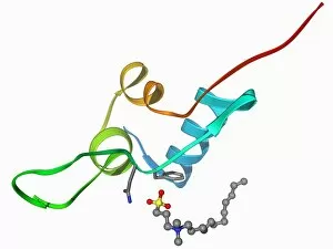
Background imageAlpha Helix Collection: Human prion precursor protein F006 / 9357

Human prion precursor protein F006 / 9357
Human prion precursor protein, molecular model showing secondary structure. The human prion protein (hPrP) is a prion precursor

Background imageAlpha Helix Collection: Serum transferrin molecule F006 / 9353

Serum transferrin molecule F006 / 9353
Serum transferrin, molecular model. Transferrins are iron-binding glycoproteins found in blood plasma

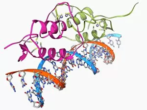
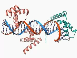
Background imageAlpha Helix Collection: Oestrogen receptor bound to DNA F006 / 9354

Oestrogen receptor bound to DNA F006 / 9354
Oestrogen receptor bound to DNA. Molecular model of the DNA-binding domain (green and pink) of the oestrogen receptor bound to a molecule of DNA (deoxyribonucleic acid, blue and orange)

Background imageAlpha Helix Collection: Human growth hormone molecule F006 / 9355

Human growth hormone molecule F006 / 9355
Human growth hormone (hGH), molecular model. hGH is produced in the anterior pituitary gland of the brain. It binds to specific receptors on cells in the body to stimulate growth

Background imageAlpha Helix Collection: Ebola matrix protein molecule F006 / 9352

Ebola matrix protein molecule F006 / 9352
Ebola matrix protein. Molecular model of the Ebola virus matrix protein VP40 bound to RNA (ribonucleic acid). This membrane-associated protein is thought to be necessary for the assembly

Background imageAlpha Helix Collection: Haemoglobin molecule F006 / 9350

Haemoglobin molecule F006 / 9350
Haemoglobin, molecular model. Haemoglobin is a metalloprotein that transports oxygen around the body in red blood cells. Each molecule consists of iron-containing haem groups (sticks)

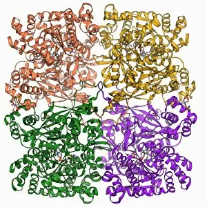
Background imageAlpha Helix Collection: Glycogen phosphorylase molecule F006 / 9347

Glycogen phosphorylase molecule F006 / 9347
Glycogen phosphorylase, molecular model. This is an enzyme involved in breaking down glycogen, the energy storage molecule involved in animal metabolism

Background imageAlpha Helix Collection: Glucose oxidase molecule F006 / 9348

Glucose oxidase molecule F006 / 9348
Glucose oxidase, molecular model. In cells this enzyme catalyses the breakdown of glucose into its metabolites

Background imageAlpha Helix Collection: Insulin-like growth 1 factor molecule F006 / 9351

Insulin-like growth 1 factor molecule F006 / 9351
Insulin-like growth factor 1 (IGF-1), molecular model. IGFs are polypeptides that are similar in their molecular structure to insulin

Background imageAlpha Helix Collection: Ribozyme enzyme and RNA F006 / 9346

Ribozyme enzyme and RNA F006 / 9346
Ribozyme enzyme and RNA, molecular model. Ribozymes are RNA (ribonucleic acid) molecules that catalyse certain biochemical reactions

Background imageAlpha Helix Collection: Transcription factors bound to DNA F006 / 9349

Transcription factors bound to DNA F006 / 9349
Transcription factors bound to DNA. Molecular model of the Oct4 (pink) and Sox2 (green) transcription factors bound to a molecule of DNA (deoxyribonucleic acid, red and blue)

Background imageAlpha Helix Collection: GA bacteriophage capsid F006 / 9341

GA bacteriophage capsid F006 / 9341
GA bacteriophage capsid, molecular model. Bacteriophages are viruses that infect bacteria, with the capsid forming the bacteriophage head


For sale as Licensed Images

Choose your image, Select your licence and Download the media

The alpha helix, a fundamental structure in biology, plays a crucial role in various molecular processes. From DNA transcription to protein synthesis, this intricate arrangement is found throughout the biological world. In the realm of genetics, the alpha helix participates in DNA transcription by aiding in the unwinding and separation of strands. Its elegant spiral shape allows for efficient reading and copying of genetic information. When it comes to proteins, the alpha helix serves as a secondary structure that contributes to their stability and function. Visualized through stunning artwork or molecular models, its coiled form adds strength and flexibility to these vital biomolecules. One example where we can observe this remarkable structure is within the nucleosome molecule. Here, DNA wraps around histone proteins forming tight coils resembling beads on a string – with each bead representing an alpha helix. Another instance occurs within bacterial ribosomes responsible for protein synthesis. The presence of multiple alpha helices enables precise positioning of molecules during translation – ensuring accurate assembly of amino acids into functional proteins. Viruses also exploit this structural motif; one such case being HIV reverse transcription enzyme. This enzyme utilizes an alpha helical region to convert viral RNA into DNA – a critical step in viral replication. Similarly, hepatitis C virus enzyme employs an intricate network of alpha helices depicted by molecular models. These structures aid in catalyzing chemical reactions necessary for viral survival and proliferation. Moving beyond viruses, manganese superoxide dismutase enzyme showcases how nature harnesses the power of the alpha helix for antioxidant defense mechanisms within cells. Its tightly wound coils protect against harmful free radicals that can damage cellular components. Alpha-helical motifs are not limited to enzymes alone but extend to larger molecules like human serum albumin or Argonaute protein involved in gene regulation pathways. Their well-defined arrangements contribute significantly to their respective functions within our bodies' complex systems.